GIDEON blog

Free Demo ► Latest Updates ► Video Tutorials ►
Subscribe , j x

Pathogen of the Month: Escherichia Coli (E. Coli)

by Dr. Jaclynn Moskow

Escherichia coli bacterium, E.coli, gram-negative rod-shaped bacteria, part of intestinal normal flora and causative agent of diarrhea and inflammations of different location, 3D illustration


Escherichia coli (E. coli) is a species of Gram-negative, rod-shaped, facultatively anaerobic bacteria. Many E. coli strains are a part of the normal flora of the gut microbiome. E. coli can also be found in the normal flora of the skin and genital tract (1).

Strains of E. coli that are part of the microbiome can be pathogenic under certain conditions – often when introduced to a new part of the body. Additionally, strains of E. coli that are not normally found in the microbiome can also cause significant disease (i.e., enterovirulent E. coli).

E. coli is the most common cause of urinary tract infection and biliary sepsis, and a common agent in travelers’ diarrhea, foodborne gastroenteritis, hemorrhagic colitis, and a wide variety of systemic infections (2).


Enterotoxigenic Escherichia Coli (ETEC)

Enterotoxigenic E. coli (ETEC) infection is the most common cause of diarrhea in children (4) and the leading cause of travelers’ diarrhea (5). It is transmitted via contaminated food and water. Symptoms commonly include watery diarrhea and abdominal cramping. Most cases are self-limited, but infection may be life-threatening in infants. 


Enteropathogenic Escherichia Coli (EPEC)

Enteropathogenic E. coli (EPEC) infection is a common cause of infantile diarrhea, although it can affect people of all ages. Like ETEC, diarrhea caused by EPEC infection is usually watery. The organism is also spread via the fecal-oral route, commonly via contaminated food and water. Infection is usually self-limited.


Uropathogenic E. Coli (UPEC)

Uropathogenic Escherichia coli (UPEC) cells adhered to bladder epithelial cell (BEC). Cells stained with methylene blue and fuchsine.
Uropathogenic Escherichia coli (UPEC) cells adhered to bladder epithelial cell (BEC). Cells stained with methylene blue and fuchsine. Author: Stefan Walkowski


E. coli strains that cause urinary tract infection are referred to as uropathogenic E. coli (UPEC). Individuals at increased risk of UPEC infection include neonates, sexually active women, geriatric individuals, and patients with indwelling urinary catheters.

Approximately 40% of adult women will experience cystitis at some point, with UPEC identified as the causative agent in 75-80% of cases (3). Untreated cystitis caused by UPEC infection can progress to pyelonephritis. Symptoms of cystitis/pyelonephritis may include dysuria, hematuria, increased urinary frequency, cloudy or foul-smelling urine, flank pain, vomiting, and fever.

Many different antibiotics are commonly used to treat UPEC infections, including penicillins, cephalosporins, fluoroquinolones, and trimethoprim-sulfamethoxazole. Treatment may be complicated by the increasing prevalence of antibiotic-resistant strains.


Shiga Toxin-Producing E. Coli (STEC)

Shiga toxin-producing E. coli (STEC) is also referred to as Verocytotoxin-producing E. coli (VTEC) or Enterohemorrhagic E. coli (EHEC). This variety of E. coli is most commonly associated with foodborne outbreaks in the developed world. Infection can be acquired from contaminated bovine meat, milk and dairy products, vegetables, fruit, and water (6).

Unlike ETEC and EPEC, infection with STEC usually causes bloody diarrhea. Treatment of diarrhea from STEC is supportive and includes fluid replacement. Infection with STEC can also cause hemolytic-uremic syndrome (HUS), most notably associated with E. coli O157:H7 strain. Nearly 40% of patients with STEC-HUS require temporary renal replacement therapy, and up to 20% will have permanent residual kidney dysfunction (2).

Worldwide, it is estimated that STEC infection causes approximately 2.8 million acute illnesses annually, 3900 cases of HUS, 270 cases of end-stage renal disease, and 230 deaths (7).

In 1993, E. coli O157:H7 made headlines when an outbreak occurred at the Jack-in-the-Box restaurant chain in the United States, affecting a total of 73 restaurant locations across 4 states. The source of this outbreak was determined to be contaminated hamburger patties. More than 700 people became ill, including 171 hospitalizations and four deaths (8). More recently, in 2019, the CDC issued a warning to avoid Romaine lettuce from the Salinas Valley region in California (9). They reported that E. coli O157:H7 infection from this vegetable affected 167 people across 27 states, with 85 hospitalizations, and 15 cases of the hemolytic uremic syndrome (10).


United States. E. coli – VTEC infection, cases and rates per 100,000
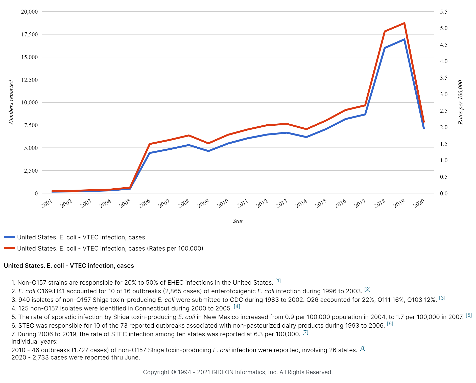
United States. E. coli - VTEC infection, cases and rates per 100,000

Want to see stats for other countries? You can, with a free trial account. Sign up here.


Enteroaggregative E. Coli (EAEC)

Enteroaggregative E. coli (EAEC) infection is recognized as the second most common cause of traveler’s diarrhea (10). It can also cause both acute and chronic childhood diarrhea. EAEC infection has been associated with reduced growth acceleration and failure to thrive among children in developing countries (11). EAEC are also the strains most commonly associated with diarrhea among individuals with HIV/AIDS (12). Diarrhea caused by EAEC is usually watery in nature. In some cases, infection is self-limiting, while in other cases, antibiotics are warranted. Fluoroquinolones, especially ciprofloxacin, are widely considered the treatments of choice (13).


Enteroinvasive E. Coli (EIEC)

Enteroinvasive E. coli (EIEC) are strains that possess some of the biochemical characteristics of E. coli and have the ability to cause dysentery through an invasion mechanism similar to that of Shigella (14).  As in shigellosis, diarrhea caused by EIEC may be watery or bloody, and mucus is sometimes present. Infection is usually self-limiting. 


Diffusely Adherent E. Coli (DAEC)

Diffusely-adherent E. coli (DAEC) is the most recent diarrheagenic E. coli pathogroup to be identified. DAEC infection is associated with diarrhea in children, where the risk of infection increases with age. These organisms have also been identified as agents of diarrhea in travelers and in patients with HIV/AIDS.  Strains have also been isolated from patients with inflammatory bowel disease and colorectal cancer (15).


Meningitis/Sepsis-Associated E. Coli (MNEC)

Meningitis/Sepsis-Associated E. coli (MNEC) infection is a common cause of severe disease in neonates. MNEC infection has a case fatality rate of 15–40% and may result in severe neurological defects in survivors (16). Third-generation cephalosporins are the recommended treatments for neonatal MNEC infection (17). Rarely, MNEC infection occurs in adults, particularly in those who are immunocompromised. 


Did you like this article? Share it on social media!

Check out more of our latest content here


References

(1) S. Baron, Medical microbiology. Galveston, Tex.: University of Texas Medical Branch at Galveston, 1996. [Online]. Available: https://www.ncbi.nlm.nih.gov/books/NBK7617/

(2) “Escherichia coli”, GIDEON Informatics, Inc, 2021. [Online]. Available: https://app.gideononline.com/explore/microbes/bacteria/escherichia-coli-1850

(3) H. Mobley, E. Hagan and M. Donnenberg, “Uropathogenic Escherichia coli”, EcoSal Plus, vol. 3, no. 2, 2009. Available: 10.1128/ecosalplus.8.6.1.3

(4) A. Mirhoseini, J. Amani and S. Nazarian, “Review on pathogenicity mechanism of enterotoxigenic Escherichia coli and vaccines against it”, Microbial Pathogenesis, vol. 117, pp. 162-169, 2018. Available: 10.1016/j.micpath.2018.02.032

(5) “Enterotoxigenic E. coli (ETEC)”, Centers for Disease Control and Prevention, National Center for Emerging and Zoonotic Infectious Diseases (NCEZID), Division of Foodborne, Waterborne, and Environmental Diseases (DFWED), 2014. [Online]. Available: https://www.cdc.gov/ecoli/etec.html

(6) “Pathogenicity assessment of Shiga toxin‐producing Escherichia coli (STEC) and the public health risk posed by contamination of food with STEC”, European Food Safety Authority, 2020. [Online]. Available: https://www.efsa.europa.eu/en/efsajournal/pub/5967

(7) Majowicz et al., “Global Incidence of Human Shiga Toxin–Producing Escherichia coliInfections and Deaths: A Systematic Review and Knowledge Synthesis”, Foodborne Pathogens and Disease, vol. 11, no. 6, pp. 447-455, 2014. Available: 10.1089/fpd.2013.1704

(8) “Jack in the Box E. Coli Outbreak – 25 Years Later”, Canadian Institute of Food Safety, 2021. [Online]. Available: https://www.foodsafety.ca/news/jack-box-e-coli-outbreak-25-years-later

(9) “The Final Update on the Multistate Outbreak of E. coli 0157:H7 Infections”, Centers for Disease Control and Prevention, 2020. [Online]. Available: https://www.cdc.gov/media/releases/2020/s0115-ecoli-outbreak.html

(10) H. Brüssow, “ESCHERICHIA COLI | Enteroaggregative E. coli”, Encyclopedia of Food Microbiology, pp. 706-712, 2014. Available: 10.1016/b978-0-12-384730-0.00387-6

(11) B. Hebbelstrup Jensen et al., “Enteroaggregative Escherichia coli in Daycare—A 1-Year Dynamic Cohort Study”, Frontiers in Cellular and Infection Microbiology, vol. 6, 2016. Available: 10.3389/fcimb.2016.00075

(12) A. Medina et al., “Diarrheagenic Escherichia coli in Human Immunodeficiency Virus (HIV) Pediatric Patients in Lima, Perú”, The American Journal of Tropical Medicine and Hygiene, vol. 83, no. 1, pp. 158-163, 2010. Available: 10.4269/ajtmh.2010.09-0596

(13) B. Hebbelstrup Jensen et al., “Characterization of Diarrheagenic Enteroaggregative Escherichia coli in Danish Adults—Antibiotic Treatment Does Not Reduce Duration of Diarrhea”, Frontiers in Cellular and Infection Microbiology, vol. 8, 2018. Available: 10.3389/fcimb.2018.00306

(14) M. Beld and F. Reubsaet, “Differentiation between Shigella, enteroinvasive Escherichia coli (EIEC) and noninvasive Escherichia coli”, European Journal of Clinical Microbiology & Infectious Diseases, vol. 31, no. 6, pp. 899-904, 2011. Available: 10.1007/s10096-011-1395-7

(15) M. Meza-Segura and T. Estrada-Garcia, “Diffusely Adherent Escherichia coli”, Escherichia coli in the Americas, pp. 125-147, 2016. Available: 10.1007/978-3-319-45092-6_6

(16) D. Wijetunge et al., “Characterizing the pathotype of neonatal meningitis causing Escherichia coli (NMEC)”, BMC Microbiology, vol. 15, no. 1, 2015. Available: 10.1186/s12866-015-0547-9

(17) Z. Zhao, X. Hua, J. Yu, H. Zhang, J. Li and Z. Li, “Duration of empirical therapy in neonatal bacterial meningitis with third-generation cephalosporin: a multicenter retrospective study”, Archives of Medical Science, vol. 15, no. 6, pp. 1482-1489, 2019. Available: 10.5114/aoms.2018.76938

World Tuberculosis Day 2021

by Dr. Jaclynn Moskow

Doctor with magnifier looking at bacteria in lungs. Tuberculosis, mycobacterium tuberculosis and world tuberculosis day concept on white background. Bright vibrant violet vector isolated illustration

Each year on March 24th, we recognize “World Tuberculosis Day” in an effort to build global awareness about the ongoing tuberculosis epidemic. Tuberculosis is an infectious disease caused by bacteria of the Mycobacterium tuberculosis complex, including Mycobacterium tuberculosis, Mycobacterium africanum, and Mycobacterium bovis (1). Worldwide, tuberculosis is the leading cause of death from an infectious agent (2).

“World Tuberculosis Day” occurs on March 24th as it was on this date, in 1884, that Dr. Robert Koch announced that he had discovered the causative agent of this disease (3).


Transmission

Tuberculosis is generally spread via the inhalation of droplet nuclei expelled by individuals with the active pulmonary or laryngeal disease. Less commonly, humans may also acquire tuberculosis from consuming unpasteurized dairy products. The incubation period of tuberculosis ranges from 4w-12w.


Clinical Manifestations

Clinical manifestations of tuberculosis vary depending on the site of mycobacterial proliferation (4). Most infections represent reactivation of a dormant focus in a lung and present with chronic fever, weight loss, nocturnal diaphoresis, and productive cough (5). Approximately 8% of patients with pulmonary tuberculosis will experience hemoptysis (6). Tuberculosis can also cause extrapulmonary disease in sites including the bone, joints, muscles, central nervous system, gastrointestinal system, hepatobiliary system, genitourinary system, eyes, breasts, and skin.

Individuals with latent tuberculosis infection (LTBI) do not experience symptoms but are carriers of the disease. They cannot spread the disease to others unless it becomes reactivated. The lifetime risk of reactivation for a person with documented LTBI is estimated to be 5–10% (7). Immunocompromised individuals are much more likely to experience tuberculosis reactivation.  


Diagnosis and Treatment

A definitive diagnosis of tuberculosis is made by the identification of the Mycobacterium tuberculosis complex in a clinical sample. Since the culture of these bacteria can be time-consuming, treatment may be initiated based on clinical suspicion alone. Tuberculosis skin tests and blood tests can be used to identify whether an individual has been infected, but cannot be used to distinguish between active and latent infections. Radiographic and other imaging techniques may also be useful in identifying patients, including those with asymptomatic active disease.


Mantoux test, positive result. Author: Grook da Oger.


Typical pulmonary infection is treated with two months of Isoniazid, Rifampin, and Pyrazinamide (with Ethambutol pending results of susceptibility testing), followed by four months of Isoniazid and Rifampin alone. Treatment of multidrug-resistant tuberculosis generally includes the use of five drugs (including Pyrazinamide and/or Rifampin) for at least 6 months, followed by four drugs for 18-24 months (5).

Patients suspected of having active tuberculosis should be isolated, and healthcare personnel should observe relevant precautions.

The Centers for Disease Control and Prevention recommends treating individuals with latent tuberculosis that are at a high risk of progressing to an active infection. Included in the “high risk” designation are individuals with HIV/AIDS and other diseases that weaken the immune system, individuals who became infected with tuberculosis in the last two years, infants and young children, the elderly, and injecting drug users (8).


Prevalence 

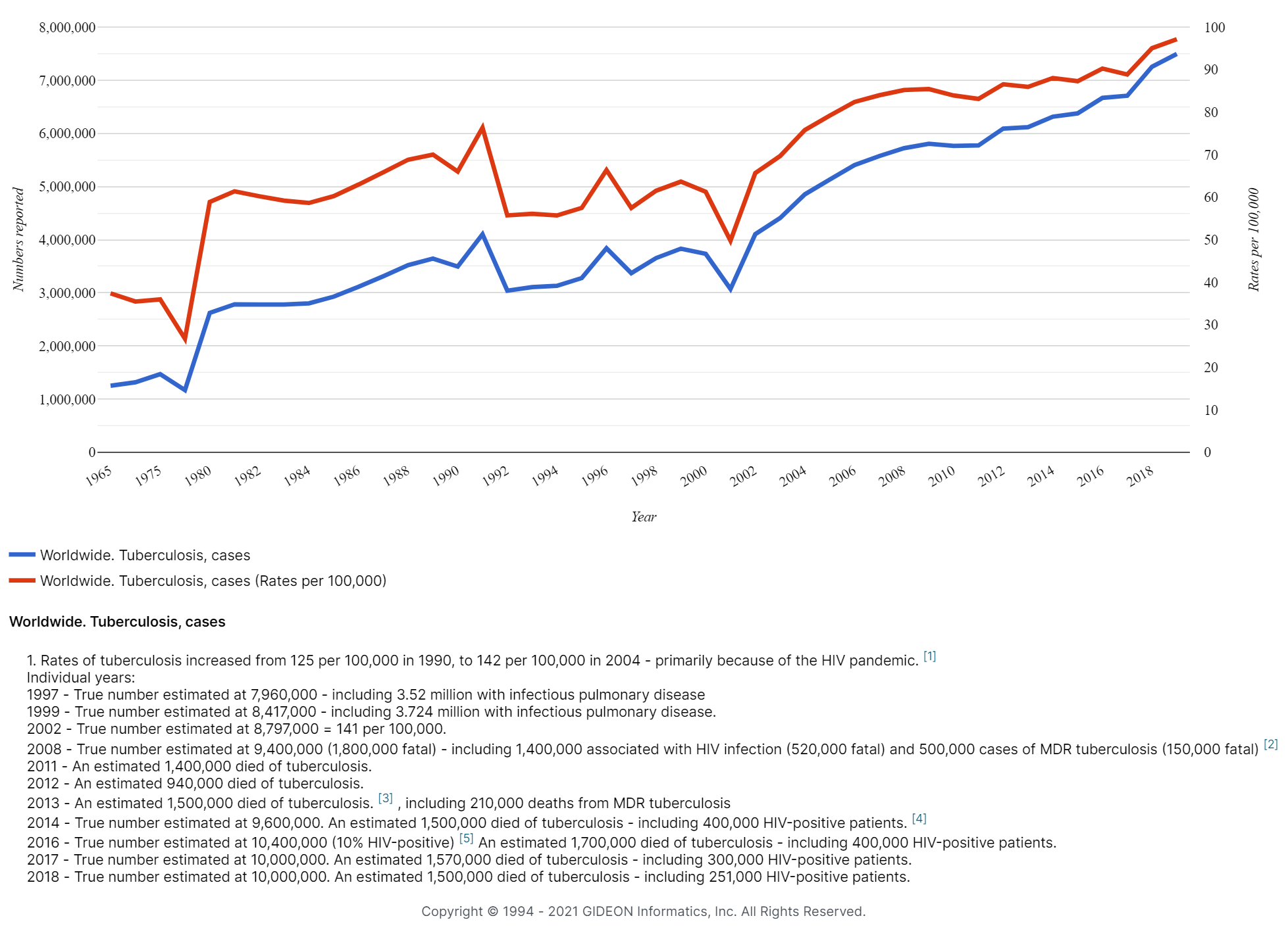
In 2018, approximately 1.7 billion individuals were infected with Mycobacterium tuberculosis – roughly 23% of the world’s population (9). In 2019, approximately 10 million individuals experienced symptomatic tuberculosis, and approximately 1.4 million died as a result of the disease (10). Tuberculosis is found worldwide, but over 95% of cases and deaths occur in developing countries (10). Eight countries currently account for two-thirds of new tuberculosis cases: India, Indonesia, China, Philippines, Pakistan, Nigeria, Bangladesh, and South Africa (10). If you have a GIDEON account, click here to explore our tuberculosis outbreak map.


Tuberculosis cases and rates Worldwide, 1965 – today

Worldwide Tuberculosis cases and rates, 1965 - today

Vaccination 

Currently, Bacille Calmette-Guerin (BCG) vaccine remains the only licensed vaccine for the prevention of tuberculosis. It provides some protection against childhood tuberculosis but is less effective in preventing adult disease (11). BCG is commonly given to children in countries in which tuberculosis is prevalent, and is estimated to decrease the risk of contracting the disease by 50% (12).


Prevention for High-Risk Travelers

The Centers for Disease Control and Prevention recommend that “travelers who anticipate possible prolonged exposure to people with tuberculosis (for example, those who expect to come in contact routinely with clinic, hospital, prison, or homeless shelter populations) should have a skin or blood test before leaving the United States. If the test reaction is negative, they should have a repeat test 8 to 10 weeks after returning to the United States. Additionally, annual testing may be recommended for those who anticipate repeated or prolonged exposure or an extended stay over a period of years.” (8)


The Future

In 2014, the World Health Organization announced that they seek to end the global tuberculosis epidemic by 2035. They defined this goal “with targets to reduce tuberculosis deaths by 95% and to cut new cases by 90%, and to ensure that no family is burdened with catastrophic expenses due to tuberculosis.”  WHO called on the cooperation and collaboration of governments, and suggested a strategy that focuses on highly vulnerable populations (such as migrants) (13). 

In 2020, they announced that the COVID-19 pandemic has stalled progress, as a result of resources being reallocated (2). They noted, for example, that many diagnostic testing machines have been used to test for COVID-19 instead of for tuberculosis. Hopefully, robust testing efforts for the disease will resume soon, as the identification of cases is critical to ending the epidemic.


Did you like this article? Share it on social media!

Check out more of our latest content here


References

(1) M. Rowe and J. Donaghy, “Mycobacterium bovis: the importance of milk and dairy products as a cause of human tuberculosis in the UK. A review of taxonomy and culture methods, with particular reference to artisanal cheeses”, International Journal of Dairy Technology, vol. 61, no. 4, pp. 317-326, 2008. Available: 10.1111/j.1471-0307.2008.00433.x

(2) “Global Tuberculosis Report”, World Health Organization, 2020. [Online]. Available: https://apps.who.int/iris/bitstream/handle/10665/336069/9789240013131-eng.pdf

(3) “The Clock Is Ticking: World TB Day 2021”, World Health Organization, 2021. [Online]. Available: https://www.who.int/campaigns/world-tb-day/world-tb-day-2021

(4) W. Cruz-Knight and L. Blake-Gumbs, “Tuberculosis”, Primary Care: Clinics in Office Practice, vol. 40, no. 3, pp. 743-756, 2013. Available: 10.1016/j.pop.2013.06.003

(5) “Tuberculosis”, GIDEON Informatics, Inc, 2021. [Online]. Available: https://app.gideononline.com/explore/diseases/tuberculosis-12470

(6) U. Seedat and F. Seedat, “Post-primary pulmonary TB haemoptysis – When there is more than meets the eye”, Respiratory Medicine Case Reports, vol. 25, pp. 96-99, 2018. Available: 10.1016/j.rmcr.2018.07.006

(7) “Latent tuberculosis infection (LTBI): FAQs”, World Health Organization, 2021. [Online]. Available: https://www.who.int/tb/areas-of-work/preventive-care/ltbi/faqs/en/

(8) “Tuberculosis: Basic TB Facts: TB Prevention”, Centers for Disease Control and Prevention, Division of Tuberculosis Elimination, 2016. [Online]. Available: https://www.cdc.gov/tb/topic/basics/tbprevention.htm

(9) “Global Health: Newsrooms: Global Health Topics: Tuberculosis”, Centers for Disease Control and Prevention, Global Health, 2020. [Online]. Available: https://www.cdc.gov/globalhealth/newsroom/topics/tb/index.html

(10) “Tuberculosis: Key Facts”, World Health Organization, 2020. [Online]. Available: https://www.who.int/news-room/fact-sheets/detail/tuberculosis

(11) S. Fatima, A. Kumari, G. Das, and V. Dwivedi, “Tuberculosis vaccine: A journey from BCG to present”, Life Sciences, vol. 252, p. 117594, 2020. Available: 10.1016/j.lfs.2020.117594

(12) G. Colditz, “Efficacy of BCG Vaccine in the Prevention of Tuberculosis”, JAMA, vol. 271, no. 9, p. 698, 1994. Available: 10.1001/jama.1994.03510330076038

(13) “WHO End TB Strategy”, World Health Organization, 2021. [Online]. Available: https://www.who.int/tb/post2015_strategy/en/

Springtime Diseases: From Spring Fever to Lyme Disease

by Dr. Jaclynn Moskow

March 20th marks the Spring Equinox when the sun crosses the equator and spring officially begins in the Northern Hemisphere. We generally associate spring with melting snow, blooming flowers, and mating animals; but did you know it is also associated with an increase in the incidence of certain diseases?

There are many factors that cause some infectious diseases to follow seasonal patterns. Changes in temperature and precipitation influence biotic and abiotic environments, disease vectors and hosts, and human behavior, including the amount of time spent outdoors (1). On a molecular level, the numbers of circulating lymphocytes and other immune cells have been observed to vary depending on the season. This may occur as a result of the circadian nature of adrenocortical hormones coupled with fluctuating vitamin D and melatonin levels (2). Additionally, temperature, moisture, and UV light can affect the infectivity of pathogens. The disease pathogens themselves, and their animal and plant reservoirs, insect vectors, and other factors ebb and flow with changes in temperature, rainfall, and many other influences. 

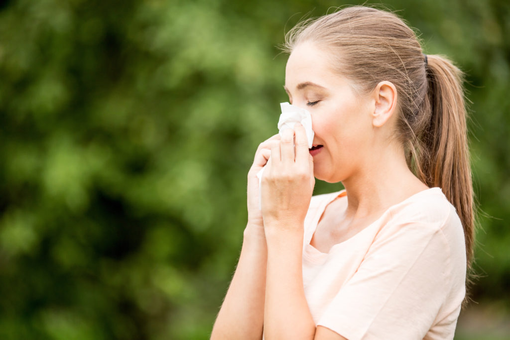
Young pretty girl blowing nose in front of blooming tree. Spring allergy concept


Spring Fever

There is actually a historical basis to the term “spring fever.” During the 18th century, individuals sometimes became ill during the springtime, experiencing weakness, joint swelling, loose teeth, and poor wound healing: the clinical manifestations of scurvy (3). As societies became more urbanized, those living in cities were faced with a lack of fruits and vegetables during the winter months, leading some to develop vitamin C deficiency.

Today, scurvy is quite rare. When seen, it is usually among alcoholics or individuals following very extreme diets (4), as opposed to city dwellers lacking access to food sources. The term “spring fever” is now used colloquially to describe a feeling of restlessness and excitement that accompanies the start of spring. “Spring fever” is a disease of the past, but other diseases of springtime remain.


Seasonal Allergies/Asthma

Allergies occur when the immune system is triggered by a non-pathogenic substance, resulting in signs and symptoms of inflammation. Many of the same substances that can trigger allergies can also trigger asthma.

Trees, grasses, and weeds produce pollen during the springtime that can instigate allergies and asthma. Additionally, certain molds that are allergenic for some people may increase in number during the spring. Individuals with allergies to pet dander may also see an increase in symptoms, as animals shed their winter coats.

Signs and symptoms of seasonal allergies include congestion, sneezing, coughing, sore throat, post-nasal drip, and headache. Eyes may become red, itchy, watery, and/or swollen. Skin rashes may also be present, as may lymphadenopathy. In extreme cases, anaphylaxis may occur. Asthma is characterized by difficulty breathing, tightness in the chest, wheezing and coughing.

It is estimated that 10–30% of the global population are affected by allergic rhinitis (5). Asthma is less common, affecting about 300 million people worldwide (6.) Those suffering from seasonal allergies will find relief by avoiding known triggers. Utilization of a HEPA filter may be of benefit, as may keeping windows and doors closed. Masks can be worn when gardening and mowing the lawn, and taking a shower immediately after these activities may also provide relief. Frequently brushing and grooming pets and vacuuming dander may also help. 

Oral antihistamines and decongestants can be used, and in extreme cases, corticosteroids may be warranted. Allergy shots are also a key option. Asthmatic attacks can be managed with a number of medications, including corticosteroids, leukotriene modifiers, beta-agonists, theophylline, ipratropium, and various immunomodulators.

Woman with a spring allergy or a cold sneezing with tissue

Rhinoviruses

Rhinoviruses are the most common causes of the common cold. Unlike influenza, which peaks in the winter, rhinovirus cases peak during the fall and spring (7). Rhinoviruses are members of the genus Enterovirus of the family Picornaviridae. Rhinovirus infection has an incubation period of 1-9 days (8).

Rhinovirus infection can resemble seasonal allergies, causing congestion, sneezing, coughing, sore throat, and headache. Unlike with seasonal allergies, muscle aches are also common, and low-grade fever may occur. Rhinoviruses can cause ear infection, and bronchiolitis/bronchitis can develop, especially in children. Rarely, pneumonia may occur. Rhinovirus may also instigate asthma attacks.

Rhinovirus infection is generally self-limiting. Patients may obtain symptomatic relief using nasal decongestants, cough suppressants, and NSAIDs. Many of the same strategies being employed to limit the spread of SARS-CoV-2 can also reduce rhinovirus transmission, including frequent hand washing, avoiding contact with those who are ill and isolating patients.


Lyme Disease

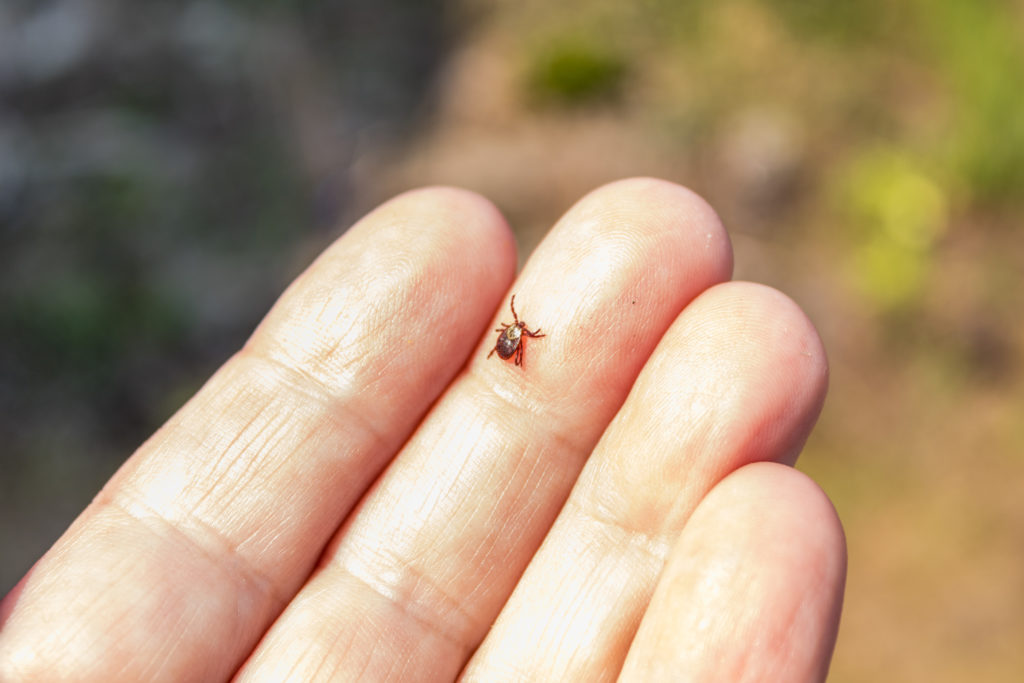
Lyme disease is caused by Borrelia spp. and transmitted to humans through the bites of infected Ixodes ticks, often referred to as black-legged ticks/deer ticks. Most cases are acquired from immature ticks (nymphs) which are small (less than 2 mm), and difficult to see. They feed during the spring and summer months (9) – the peak season of Lyme disease (10).

The tick Ixodes ricinus crawling on human skin. This kind of animal is a distributor of Borrelia spp, an agent of Lyme disease
Ixodes ricinus tick is a distributor of Borrelia spp., an agent of Lyme disease


Lyme disease has an incubation period ranging from 2-180 days, with most cases manifesting within 7 to 14 days. About 25% of patients recall a recent tick bite. Erythema migrans is present in 75% of cases and is usually neither pruritic nor painful. Multiple skin lesions may occur in 20% to 50% of cases. A nodule in the nipple or ear lobe (borrelial lymphocytoma) may be present. Acrodermatitis chronicum atrophicans can also occur, typically seen on the hands and feet (11). 

Neurological manifestations occur in 10-15% of patients (12). The most common of these include lymphocytic meningitis, cranial neuritis, mononeuropathy multiplex, and painful radiculoneuritis. The range of joint involvement includes tendonitis, myositis, and bursitis, which wax and wane. The cardiac disease may be characterized by arrhythmia, heart block, chest pain, and pericarditis or myopericarditis. Rarely, other organs may become involved. 

Doxycycline, Ceftriaxone, Amoxicillin, and Cefuroxime can be used as a treatment, with dosage, route, and duration varying according to patient age and the nature and severity of the disease.

About 30,000 cases of Lyme Disease are reported to the Centers for Disease Control and Prevention (CDC) each year, but they estimate that as many as 476,000 people will actually contract the disease (13). Most cases occur in Pennsylvania, New York, Connecticut, and other states in the Northeastern United States. The disease is also common in Wisconsin and Minnesota. Lyme disease has been reported in Asia: in China, Korea, Japan, Indonesia, Nepal, and eastern Turkey. In Europe, most Lyme disease cases occur in Scandinavian countries, Germany, Austria, and Slovenia (14).

The CDC recommends that individuals spending time in wooded and grassy areas perform daily “tick checks.” By removing a tick within 24 hours, Lyme disease transmission is greatly decreased. It is important to contact a health professional before attempting to remove a tick. When outdoors, covering skin by wearing long clothing can also reduce transmission. 

Stay safe and Happy Spring!


Did you like this article? Share it on social media!

Check out more of our latest content here


References

(1) M. Martinez, “The calendar of epidemics: Seasonal cycles of infectious diseases”, PLOS Pathogens, vol. 14, no. 11, p. e1007327, 2018. Available: 10.1371/journal.ppat.1007327

(2) A Fares A, “Factors influencing the seasonal patterns of infectious diseases”, Int J Prev Med, vol. 4, no. 2, pp. 128-32, 2013.

(3) P. Janson, “When Spring Fever Was a Real Disease”, Emergency Medicine News, vol. 38, p. 1, 2016. Available: 10.1097/01.eem.0000484361.70086.35

(4) M. Weinstein, P. Babyn and S. Zlotkin, “An Orange a Day Keeps the Doctor Away: Scurvy in the Year 2000”, PEDIATRICS, vol. 108, no. 3, pp. e55-e55, 2001. Available: 10.1542/peds.108.3.e55

(5) C. Schmidt, “Pollen Overload: Seasonal Allergies in a Changing Climate”, Environmental Health Perspectives, vol. 124, no. 4, 2016. Available: 10.1289/ehp.124-a70

(6) “Asthma”, Who.int, 2021. [Online]. Available: https://www.who.int/news-room/fact-sheets/detail/asthma

(7) A. Monto, “The seasonality of rhinovirus infections and its implications for clinical recognition”, Clinical Therapeutics, vol. 24, no. 12, pp. 1987-1997, 2002. Available: 10.1016/s0149-2918(02)80093-5

(8) Lessler, N. Reich, R. Brookmeyer, T. Perl, K. Nelson and D. Cummings, “Incubation periods of acute respiratory viral infections: a systematic review”, The Lancet Infectious Diseases, vol. 9, no. 5, pp. 291-300, 2009. Available: 10.1016/s1473-3099(09)70069-6

(9) “Lyme disease: Transmission”, Centers for Disease Control and Prevention, National Center for Emerging and Zoonotic Infectious Diseases (NCEZID), Division of Vector-Borne Diseases (DVBD), 2020. [Online]. Available: https://www.cdc.gov/lyme/transmission/index.html

(10) S. Moore, R. Eisen, A. Monaghan and P. Mead, “Meteorological Influences on the Seasonality of Lyme Disease in the United States”, The American Journal of Tropical Medicine and Hygiene, vol. 90, no. 3, pp. 486-496, 2014. Available: 10.4269/ajtmh.13-0180

(11) “Lyme disease”, GIDEON Informatics, Inc, 2021. [Online]. Available: https://app.gideononline.com/explore/diseases/lyme-disease-11360

(12) J. Halperin, “Neurologic Manifestations of Lyme Disease”, Current Infectious Disease Reports, vol. 13, no. 4, pp. 360-366, 2011. Available: 10.1007/s11908-011-0184-x

(13) “Lyme disease: Data and Surveillance”, Centers for Disease Control and Prevention, National Center for Emerging and Zoonotic Infectious Diseases (NCEZID), Division of Vector-Borne Diseases (DVBD), 2021. [Online]. Available: https://www.cdc.gov/lyme/datasurveillance/index.html?CDC_AA_refVal=https%3A%2F%2Fwww.cdc.gov%2Flyme%2Fstats%2Findex.html

(14) Meyerhoff, J, “What is the global prevalence of Lyme disease?”, Medscape.com, 2019. [Online]. Available: https://www.medscape.com/answers/330178-101008/what-is-the-global-prevalence-of-lyme-disease

Reviewing Fungal Infections

by Dr. Jaclynn Moskow

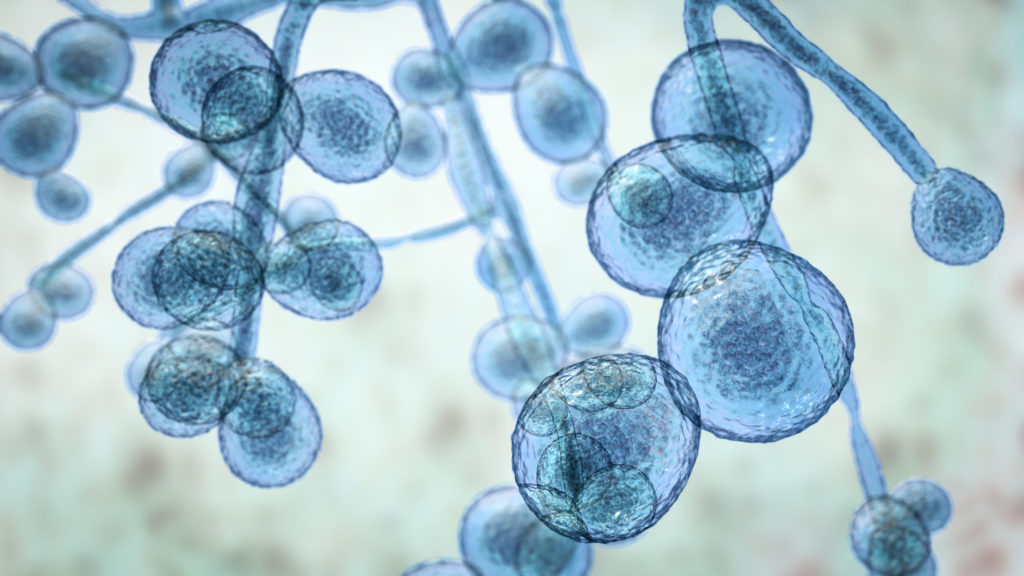
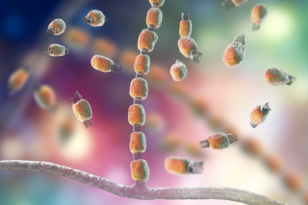
Candida auris fungi, emerging multidrug resistant fungus, agent of fungal infection
Candida auris, an emerging multidrug-resistant fungus


Fungi are similar in many ways to bacteria – both have a cell nucleus and complex cell walls. Unlike bacteria, species of fungi include both single-celled organisms (yeasts) and multicellular forms (molds). Molds resemble plants and often consist of filaments, spores, root structures, etc. Fungal infections (mycoses) include candidiasis, dermatophytosis, blastomycosis, coccidioidomycosis, histoplasmosis, cryptococcosis, paracoccidioidomycosis, aspergillosis, zygomycosis, and pneumocystosis.


Candidiasis

Candidiasis refers to infections caused by yeasts of the genus Candida. Candida is the most common cause of fungal infections worldwide; and is part of the normal flora of the mouth, GI tract, vagina, and skin. Candidiasis occurs when an imbalance in the amount of Candida in these areas results in signs and symptoms of inflammation, or when Candida colonizes parts of the body in which it is not normally present. All forms of candidiasis are more common in individuals who are immunocompromised. 

Vulvovaginal candidiasis fungal infection, commonly referred to as a “yeast infection,” is estimated to affect 70-75% of women at least once during their lifetimes (1). Symptoms may include itching, burning, soreness, redness, swelling, pain during intercourse or urination, and a thick, white discharge that is usually odorless and may resemble cottage cheese. Factors that predispose to vulvovaginal candidiasis include the use of antibiotics, douches, and other vaginal products, diabetes, hormonal changes such as those seen with pregnancy and menopause, contraceptives, immune deficiency, including HIV / AIDS, and certain genetic factors. A variety of topical and systemic azole agents can be used for treatment.

Oropharyngeal candidiasis commonly referred to as “thrush,” occurs from Candida overgrowth on the lining of the mouth, tongue, gums, tonsils, and lips. The condition may cause visible white or yellow patches, soreness, an unpleasant taste, and occasionally a “cotton-like sensation.” It is much more common in infants and toddlers than in adults. Predisposing factors in adults include smoking, dentures, antibiotic and corticosteroid use, and hormonal changes. 80-90% of HIV patients will experience oropharyngeal candidiasis (2). Proper dental hygiene may help protect against oropharyngeal candidiasis. Various azole mouthwashes, gels, and lozenges can be used for treatment, as well as oral antifungal medications.

Common sites of cutaneous candidiasis include the axilla (armpit), the area under the breast, the groin region, the intergluteal cleft, and on the hands and feet. Candida is a common cause of “diaper rash.”

Invasive candidiasis (“deep candidiasis”) occurs when Candida affects the bloodstream, heart, brain, eyes, bones, or other organs. It may occur in patients that are immunocompromised, or as a result of fungal infection introduced by vascular lines, prosthetic cardiac valves, and urinary catheters. Systemic symptoms may result, including fever, chills, pain, hypotension, and neurological deficits. The condition can be fatal. One strain, in particular, Candida auris, poses a threat in hospitals, as it is often multidrug-resistant and difficult to identify using standard laboratory methods (3).


Dermatophytosis

Dermatophytosis (“tinea”) is a fungal infection of keratinized tissue, including the skin, hair, and nails. Fungal causes include Ascomycota, Euascomycetes, Onygenales: Epidermophyton, Microsporum, Trichophyton, Trichosporon spp., and Arthroderma (4). Dermatophytosis is contracted by contact with infected humans or animals, or contact with contaminated objects, flooring, or soil.

Trichophyton mentagrophytes - an agent of fungal infection
Fungus Trichophyton mentagrophytes


The nomenclature of these conditions derives from the body region that is affected. For example, Tinea manuum is a dermatophyte infection of the hands, while Tinea barbae is an infection of the beard or mustache. Tinea pedis affects the feet, Tinea unguium the nails, Tinea cruris the groin, Tinea corporis the trunk, Tinea capitis the scalp, and Tinea faciei the non-bearded area of the face. 

Tinea corporis is commonly referred to as “ringworm.” It presents as a red, annular, scaly patch, often with central clearing. The condition is usually pruritic and is very common – especially among children. High rates are seen in Africa, India, and urban areas of the Americas (5). A common source of adult infection is through handling puppies and kittens. A wide variety of creams, ointments, gels, and sprays are available for treatment.

Tinea pedis is commonly referred to as “athlete’s foot”; and is the most common form of dermatophytosis in adults (6). The condition can cause itching, stinging, and burning of the feet – often with redness, blisters, and peeling. Tinea pedis is often acquired from wet floor surfaces such as showers, locker rooms, and pool areas. Wearing foot protection in these areas can help prevent transmission. 

The same fungal species that cause Tinea pedis can also cause Tinea cruris, commonly known as “jock itch.” Tinea cruris presents as a red, pruritic, and often annular rash in the crease of the groin. The condition may spread to the upper thigh in a “half-moon” shape. The condition can be acquired by sharing contaminated towels or clothing. Both Tinea pedis and Tinea cruris usually respond well to topical antifungals.


Endemic Mycoses

Endemic mycoses refer to a diverse group of fungal infections found in distinct geographical regions. They can cause significant morbidity and mortality in immunocompromised individuals, and may also affect healthy people. 

Blastomycosis is caused by the fungus Blastomyces. It mainly affects people living in regions of the United States and Canada surrounding the Ohio and Mississippi River valleys and the Great Lakes (7). Blastomycosis is acquired through inhalation of spores, often after participating in activities that disturb the soil. Symptoms are “flu-like” and may include fever, fatigue, muscle aches, night sweats, and cough. A chronic disease may affect the lungs, skin, bones, joints, genitourinary tract, or central nervous system. Amphotericin B is the treatment of choice.

Coccidioidomycosis (“Valley Fever”) is caused by Coccidioides immitis and Coccidioides posadasii. The condition is found in the Southwestern United States and parts of Mexico and Central and South America. Like blastomycosis, coccidioidomycosis follows the inhalation of spores from the soil. Symptoms are similar to coccidioidomycosis and are flu-like. A rash on the upper body or legs is commonly encountered. Most people with coccidioidomycosis improve without treatment, but fluconazole and similar antifungals can be used (8).

Fungus Coccidioides immitis, saprophytic stage, 3D illustration showing fungal arthroconidia. Pathogenic fungi that reside in soil and can cause fungal infection Coccidioidomycosis, or Valley fever
Coccidioides immitis, an agent of fungal infection Coccidioidomycosis (aka Valley fever), saprophytic stage.


Histoplasmosis, caused by Histoplasma, is acquired by inhaling spores – usually from soil containing bird- or bat-droppings.  The condition is found in the Ohio and Mississippi River valleys and parts of Central and South America, Africa, Asia, and Australia (9).  Histoplasmosis is also characterized by a flu-like illness and is usually self-limiting. 

Cryptococcosis is caused by various species of Cryptococcus, yeasts that are found in the soil and on certain trees. Cryptococcus gattii is found in California, Oregon, Washington, Canada, Australia, Papua New Guinea, and South America (10). Cryptococcus neoformans is found in all countries. Cryptococcus is often associated with pneumonia or meningitis. The current global incidence is estimated at 1 million cases per year, with 50% mortality (11). Most of these cases occur in individuals with HIV / AIDS. Treatment consists of Amphotericin B and Flucytosine, followed by Fluconazole.

Paracoccidioidomycosis is caused by Paracoccidioides, found in parts of Central and South America (12). It can cause lesions in the mouth and throat, rash, lymphadenopathy, fever, cough, and hepatosplenomegaly. Talaromycosis, formally known as sporotrichosis, is an endemic mycosis caused by Talaromyces marneffei and other species. The condition is found in Southeast Asia, Southern China, and Eastern India (13). Clinical manifestations include fever, cough, lymphadenopathy, hepatosplenomegaly, diarrhea, and abdominal pain.


Mold Infections

Most people inhale mold spores every day without becoming ill, but occasionally severe disease can result. Infection by Aspergillus (aspergillosis) may present as an allergic reaction. The fungus can also cause infection of the sinuses and lungs. Formation of “fungal ball” (aspergillomas) may occur in patients with pre-existing lung diseases. Aspergillus can also infect the eyes, skin, cardiac valves, brain, gastrointestinal tract, and genitourinary tract. Treatment options include Voriconazole, Amphotericin B, and Isavuconazole (14).

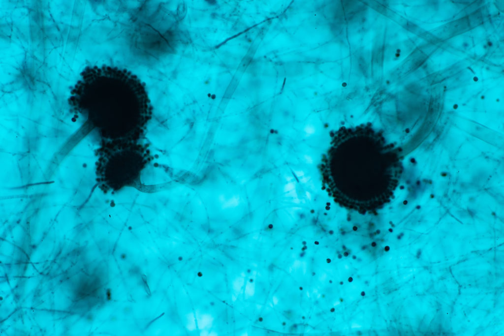
Aspergillus (mold) under the microscopic view. Aspergillus is an agent of fungal infection.
Aspergillus spp. under a microscope


Zygomycosis (“mucormycosis”) is caused by a group of molds called Mucormycetes. This fungal infection is commonly associated with hyperglycemia, metabolic (diabetic, uremic) acidosis, corticosteroid therapy, and neutropenia, transplantation, heroin injection, and administration of deferoxamine (15). Common sites of infection include the paranasal sinuses and contiguous structures, cranial nerves, cerebral arteries, lungs, and skin. Treatment may include intravenous Amphotericin B, followed by oral Posaconazole or Isavuconazole.

Other molds that can cause allergies and infections in humans include Stachybotrys chartarum, Alternaria alternata, Lomentospora prolificans, Scedosporium apiospermum, Cladosporium, and Penicillium.

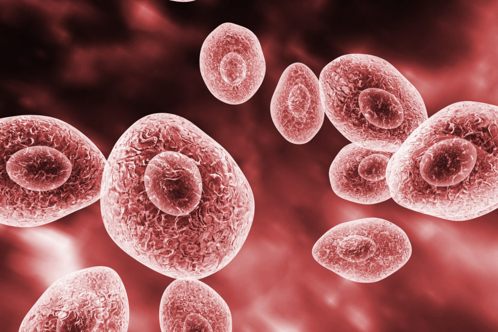
Pneumocystis jirovecii, an agent of Pneymocystis pneumonia
Pneumocystis jirovecii, an agent of Pneumocystis pneumonia fungal infection

Pneumocystis pneumonia

Pneumocystis pneumonia (PCP) is caused by the fungus Pneumocystis jirovecii.  Until recent years, the organism had been classified as a protozoan parasite. Pneumocystis pneumonia usually occurs in individuals with severe immune suppression, including HIV / AIDS.  Presenting symptoms include shortness of breath, fever, and a nonproductive cough. Extrapulmonary infection is rare but can occur. Treatment options include Sulfamethoxazole / Trimethoprim, Pentamidine, Dapsone + Trimethoprim, Atovaquone, or Primaquine + Clindamycin (16).


Did you like this article? Share it on social media!

Check out more of our latest content here


References

(1) Sobel JD. Vulvovaginal candidosis. Lancet. 2007 Jun 9;369(9577):1961-71. doi: 10.1016/S0140-6736(07)60917-9.

(2) Patil S, Majumdar B, Sarode SC, Sarode GS, Awan KH. Oropharyngeal Candidosis in HIV-Infected Patients-An Update. Front Microbiol. 2018 May 15;9:980. doi: 10.3389/fmicb.2018.00980.

(3) “General Information about Candida auris”, Centers for Disease Control and Prevention, National Center for Emerging and Zoonotic Infectious Diseases (NCEZID), Division of Foodborne, Waterborne, and Environmental Diseases (DFWED), 2019. [Online]. Available: https://www.cdc.gov/fungal/candida-auris/candida-auris-qanda.html

(4)”Dermatophytosis”, GIDEON Informatics, Inc, 2021. [Online]. Available: https://app.gideononline.com/explore/diseases/dermatophytosis-10600

(5) M. Handler, “What is the global incidence of tinea capitis (scalp ringworm)?”, Medscape.com, 2020. [Online]. Available: https://www.medscape.com/answers/1091351-36134/what-is-the-global-incidence-of-tinea-capitis-scalp-ringworm

(6) Ilkit M, Durdu M. Tinea pedis: the etiology and global epidemiology of a common fungal infection. Crit Rev Microbiol. 2015;41(3):374-88. doi: 10.3109/1040841X.2013.856853.

(7) “Blastomycosis”, Centers for Disease Control and Prevention, National Center for Emerging and Zoonotic Infectious Diseases (NCEZID), Division of Foodborne, Waterborne, and Environmental Diseases (DFWED), 2020. [Online]. Available: https://www.cdc.gov/fungal/diseases/blastomycosis/index.html

(8) “Treatment for Valley Fever (Coccidioidomycosis)”, Centers for Disease Control and Prevention, National Center for Emerging and Zoonotic Infectious Diseases (NCEZID), Division of Foodborne, Waterborne, and Environmental Diseases (DFWED), 2019. [Online]. Available: https://www.cdc.gov/fungal/diseases/coccidioidomycosis/treatment.html

(9) “Histoplasmosis”, Centers for Disease Control and Prevention, National Center for Emerging and Zoonotic Infectious Diseases (NCEZID), Division of Foodborne, Waterborne, and Environmental Diseases (DFWED), 2020. [Online]. Available: https://www.cdc.gov/fungal/diseases/histoplasmosis/index.html

(10) “C. gattii Infection Statistics”, Centers for Disease Control and Prevention, National Center for Emerging and Zoonotic Infectious Diseases (NCEZID), Division of Foodborne, Waterborne, and Environmental Diseases (DFWED), 2020. [Online]. Available: https://www.cdc.gov/fungal/diseases/cryptococcosis-gattii/statistics.html

(11)”Cryptococcosis worldwide distribution”, GIDEON Informatics, Inc, 2021. [Online]. Available: https://app.gideononline.com/explore/diseases/cryptococcosis-10530/worldwide

(12) “Paracoccidioidomycosis”, Centers for Disease Control and Prevention, National Center for Emerging and Zoonotic Infectious Diseases (NCEZID), Division of Foodborne, Waterborne, and Environmental Diseases (DFWED), 2020. [Online]. Available: https://www.cdc.gov/fungal/diseases/other/paracoccidioidomycosis.html

(13) “Talaromycosis (formerly Penicilliosis)”, Centers for Disease Control and Prevention, National Center for Emerging and Zoonotic Infectious Diseases (NCEZID), Division of Foodborne, Waterborne, and Environmental Diseases (DFWED), 2020. [Online]. Available: https://www.cdc.gov/fungal/diseases/other/talaromycosis.html

(14) “Aspergillosis”, GIDEON Informatics, Inc, 2021. [Online]. Available: https://app.gideononline.com/explore/diseases/aspergillosis-10140

(15) “Zygomycosis”, GIDEON Informatics, Inc, 2021. [Online]. Available: https://app.gideononline.com/explore/diseases/zygomycosis-12670

(16) “Pneumocystis pneumonia”, GIDEON Informatics, Inc, 2021. [Online]. Available: https://app.gideononline.com/explore/diseases/pneumocystis-pneumonia-11850

Congenital Infections: TORCH

by Dr. Jaclynn Moskow

Pregnant woman cuddling her belly
By obtaining proper prenatal and perinatal care, pregnant women can optimize their chances of preventing, detecting, and treating congenital infections.


Congenital infections are caused by pathogens transmitted from a mother to her offspring during pregnancy or delivery. These infections can cause significant fetal and neonatal morbidity and mortality. The mnemonic “TORCH” is often used to refer to common congenital infections:


T – Toxoplasma

O – Other (Syphilis, Parvovirus B19, Varicella-Zoster, etc)

R – Rubella

C – CMV

H – Herpes Simplex Virus


Congenital Toxoplasmosis

Congenital toxoplasmosis is caused by the parasite Toxoplasma gondii. It can be acquired when a pregnant woman consumes raw or undercooked meat, or contacts contaminated water, soil, or cat feces (generally from outdoor cats that hunt.) The classic triad of congenital toxoplasmosis is 1) chorioretinitis 2) hydrocephalus and 3) cerebral calcifications. 

Symptoms often do not occur until months after birth and may include seizures, cognitive impairment, and cerebellar dysfunction (1). The retinal disease associated with congenital toxoplasmosis is progressive. Other clinical manifestations may include fever, rash, hydrocephalus or microcephaly, sensorineural hearing loss, congenital nephro­sis, hematologic abnormalities, hepatosplenomegaly, various endocrinopathies, and myocarditis. Infection can lead to spontaneous abortion, prematurity, stillbirth, and perinatal death. 

Congenital toxoplasmosis is more severe when acquired in early pregnancy. The incidence is highest in the Eastern Mediterranean and Africa (2). Rates are estimated at 1 per 3000-10000 live births in the United States (3) and 6.7 per 10000 live births in Europe – with 81% of all confirmed cases in the EU/EEA occurring in France (4). Spiramycin can decrease the risk of vertical transmission, but will not treat the fetus if the infection has already occurred. Infants born with this disease may benefit from pyrimethamine, sulfadiazine, and leucovorin. 

To help prevent infection, pregnant women should avoid consuming raw and undercooked meat, wear gloves when gardening, and avoid changing cat litter.


Congenital Rubella

Rubella, also known as “German measles,” is caused by the Rubella virus (Togaviridae family). It is most severe when acquired during the first trimester of pregnancy when the maternal infection will lead to fetal demise in 40-90% of cases (5). Congenital rubella can cause cardiac abnormalities, including patent ductus arteriosus and pulmonary artery stenosis. It can also cause ophthalmic abnormalities such as cataracts, glaucoma, retinopathy, and microphthalmia. Sensorineural deafness is common, and microcephaly, cognitive impairment, and meningoencephalitis may occur. Hepatosplenomegaly, hepatitis, hemolytic anemia, and thrombocytopenic purpura may also be observed. 

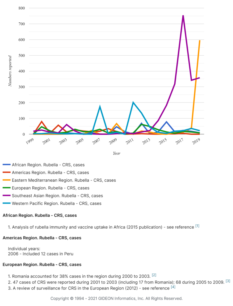
The incidence of congenital rubella has plummeted in countries that employ widespread vaccination. In recent times, documented cases of rubella in the United States are virtually all imported. Cases and outbreaks continue in Europe but at a very low rate. In 2008, 48% of all cases occurred in Southeast Asia and 38% in Africa (6). There is no effective treatment for congenital rubella.


Regional comparison of Congenital Rubella Syndrome prevalence, 1999 – 2019

Congenital infections - Congenital Rubella Syndrome prevalence: comparison between regions worldwide, 1999 - 2019


Congenital CMV

Congenital cytomegalovirus (CMV) is the most common congenital viral infection in the developed world. Clinical manifestations include sensorineural hearing loss, visual impairment, cerebral palsy, and cognitive difficulty. It can also cause neonatal cholestasis, pulmonary hypertension, and epilepsy. 10-20% of all hearing impairment in children is caused by congenital CMV (7).

CMV is identified in 5 to 7 per 1000 live births in the USA, Canada, Western Europe, and Australia; and 10-30 per 1000 live births in Latin America, Africa, and most Asian countries (8). Symptomatic infants may benefit from treatment with valganciclovir. It is difficult to prevent the acquisition of CMV, but some have suggested that pregnant women can decrease risk by avoiding contact with the saliva and urine of young children.


Congenital HSV

Congenital herpes simplex virus (HSV) most commonly occurs when an infant is exposed to the mother’s genital tract during delivery. Both herpes simplex-1 and herpes simplex-2 can cause congenital HSV. The risk of transmission from mother to infant depends primarily on when the maternal infection was acquired. When a mother is infected close to the time of delivery, the fetal infection rate is estimated at 25-60%. This rate drops to less than 2% when a mother is infected during the first half of pregnancy or earlier (9).

Signs of congenital HSV infection may occur between birth and six weeks of age. Disseminated disease may involve the liver, lung, central nervous system, and skin. “SEM disease” is limited to the skin, eyes, and/or mouth. Congenital HSV may cause a vesicular rash, hypothermia, lethargy, seizures, respiratory distress, hepatosplenomegaly, thrombocytopenia, hepatic dysfunction, cerebrospinal fluid pleocytosis, and sepsis. Congenital HSV is fatal in 50% of cases (10). The incidence of congenital HSV is estimated to be between 1 in 3000-20000 live births. All pregnant women should be tested for HSV, and those who are positive should receive prophylactic acyclovir or a similar drug at the time of delivery. Infected infants should be treated as well.


Congenital Syphilis

Congenital syphilis occurs when the bacterium Treponema pallidum is transmitted transplacentally or via the birth canal. The rate of vertical transmission increases as the pregnancy advances and transmission is more likely when the mother is experiencing early disease (11). Congenital syphilis can sometimes be detected by the appearance of nonimmune hydrops fetalis on ultrasound examination.

Congenital syphilis may be divided into two clinical syndromes: early congenital syphilis and late congenital syphilis. The early disease manifests within the first two years of life and is characterized by rash, adenopathy, and hepatosplenomegaly. Mucous patches and condylomata lata may be seen.  The eyes may be affected, and cranial nerve palsy and seizures may occur. Thrombocytopenia with petechiae and purpura are often noted. Other manifestations can include anemia, myocarditis, pancreatitis, nephrotic syndrome, and malabsorption. Osteochondritis is often seen on imaging. 

Late congenital syphilis manifests after two years of age. Dental findings include “Hutchinson’s teeth” and “mulberry molars.” Interstitial keratitis and eighth cranial nerve deafness can occur. Rhagades may be seen. Bone and joint abnormalities may include frontal bossing, saddle nose deformity, protuberant mandible, short maxilla, high palatal arch, sternoclavicular joint thickening (Higouménakis sign), saber shin, and Clutton’s joints. Central nervous system involvement can include cognitive impairment, hydrocephalus, seizures, cranial nerve palsy, paralysis, and optic nerve atrophy.


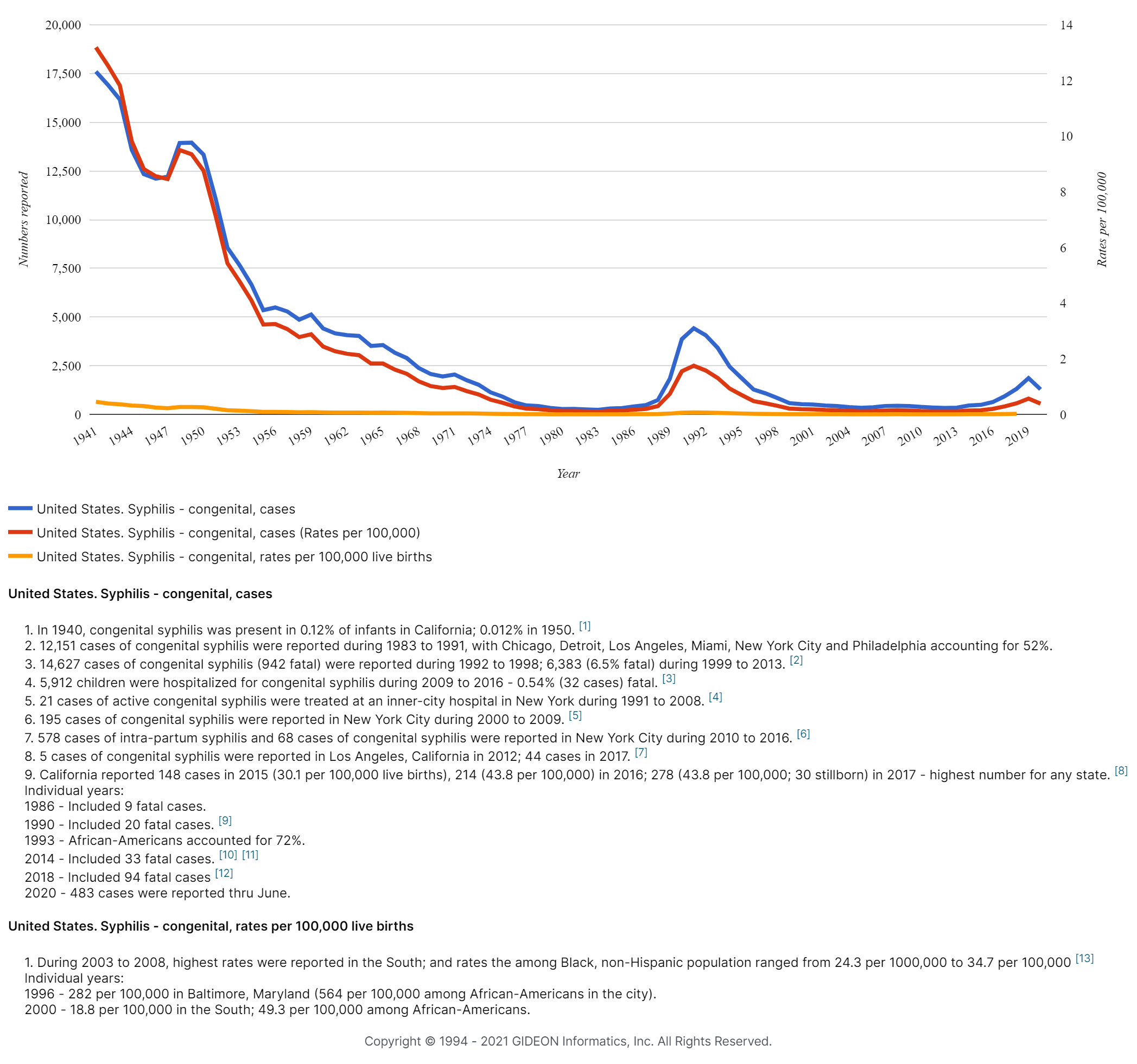
Congenital infections, Syphilis in the United States, 1941 – 2019


Graph illustrating the prevalence of congenital infection - Syphilis in the United States, 1941 - 2019


The WHO estimates that there were approximately 661,000 total cases of congenital syphilis in 2016, resulting in over 200,000 stillbirths and neonatal deaths (12) – with most cases occurring in South America and Africa. The CDC reports that congenital syphilis is on the rise in the United States, with the number of cases in 2018 being highest since 1998 (13).

All pregnant women should be tested for syphilis at their first prenatal visit. Penicillin is the only known effective antimicrobial agent for the prevention of vertical transmission and treatment of fetal and neonatal infection.


Congenital Parvovirus B19

Parvovirus B19 is estimated to infect 1-5% of pregnant women. Most infections are without consequence to the fetus, but in rare cases, serious fetal disease can arise (14). In infected fetuses, ultrasound may show nonimmune hydrops fetalis. 

Congenital parvovirus B19 often causes severe anemia and may also cause thrombocytopenia. Neurological manifestations include hydrocephalus, cerebellar hemorrhage, and polymicrogyria. Cardiac complications can include Ebstein’s anomaly, ventricular septal defect, cardiomyopathy, second‐degree heart block, and myocarditis. Ocular involvement may include corneal opacification, aphakia, and microphthalmia. Gastrointestinal manifestations include meconium peritonitis, fetal liver calcifications, portal tract fibrosis, and hypoplasia of the abdominal muscles. Congenital parvovirus B19 can also cause cleft lip and palate, micrognathia, bifid scrotum, secundum atrial septal defect, and micropenis with perineoscrotal hypospadias.

Intrauterine fetal blood transfusion can be used to treat the severe fetal anemia associated with congenital parvovirus B19 infection. 


Congenital Varicella-Zoster

Varicella-zoster congenital infections are caused by the virus that causes chickenpox and shingles. Infection may be characterized by low birth weight, hypoplasia of the extremities, dermal scarring, focal muscular atrophy, encephalitis, cortical atrophy, chorioretinitis, and microcephaly. Neonatal varicella zoster may occur when a mother contracts varicella virus between five days before delivery – to 48 hours after delivery. Neonatal varicella has a fatality rate of up to 30% (15). Congenital Varicella-Zoster virus infection is rare since most women are immune by childbearing age – having either been infected during childhood or vaccinated. Infants born with congenital varicella zoster may improve with acyclovir. 


Additional Congenital Infections

Additional viral agents of fetal and neonatal morbidity and mortality include HIV, Hepatitis B and C, measles, enteroviruses, adenovirus, lymphocytic choriomeningitis virus, West Nile virus, Zika virus, and Chikungunya virus. Additional bacterial causes include Group B Streptococcus, Chlamydia trachomatis, Neisseria gonorrhoeae, , Escherichia coli, Mycobacterium tuberculosis, and Coxiella burnetii. A parasite, Plasmodium falciparum (the causative agent of malaria) is also associated with congenital infection. 

By obtaining proper prenatal and perinatal care, pregnant women can optimize their chances of preventing, detecting, and treating congenital infections. 


Did you like this article? Share it on social media!

Check out more of our latest content here


References

(1) Hampton MM. Congenital Toxoplasmosis: A Review. Neonatal Netw. 2015;34(5):274-8. doi: 10.1891/0730-0832.34.5.274.

(2) Rostami A, Riahi SM, Contopoulos-Ioannidis DG, Gamble HR, Fakhri Y, Shiadeh MN, Foroutan M, Behniafar H, Taghipour A, Maldonado YA, Mokdad AH, Gasser RB. Acute Toxoplasma infection in pregnant women worldwide: A systematic review and meta-analysis. PLoS Negl Trop Dis. 2019 Oct 14;13(10):e0007807. doi: 10.1371/journal.pntd.0007807.

(3) McAuley JB. Congenital Toxoplasmosis. J Pediatric Infect Dis Soc. 2014 Sep;3 Suppl 1(Suppl 1):S30-5. doi: 10.1093/jpids/piu077.

(4) “Congenital toxoplasmosis – Annual Epidemiological Report for 2016”, European Centre for Disease Prevention and Control, 2021. [Online]. Available: https://www.ecdc.europa.eu/en/publications-data/congenital-toxoplasmosis-annual-epidemiological-report-2016. 

(5) Best JM. Rubella. Semin Fetal Neonatal Med. 2007 Jun;12(3):182-92. doi: 10.1016/j.siny.2007.01.017. 

(6) Bouthry E, Picone O, Hamdi G, Grangeot-Keros L, Ayoubi JM, Vauloup-Fellous C. Rubella and pregnancy: diagnosis, management and outcomes. Prenat Diagn. 2014 Dec;34(13):1246-53. doi: 10.1002/pd.4467. 

(7) Goderis J, De Leenheer E, Smets K, Van Hoecke H, Keymeulen A, Dhooge I. Hearing loss and congenital CMV infection: a systematic review. Pediatrics. 2014 Nov;134(5):972-82. doi: 10.1542/peds.2014-1173. 

(8) Fowler KB, Boppana SB. Congenital cytomegalovirus infection. Semin Perinatol. 2018 Apr;42(3):149-154. doi: 10.1053/j.semperi.2018.02.002.

(9) Fernandes ND, Arya K, Ward R. Congenital Herpes Simplex. 2021 Jan 11. In: StatPearls [Internet]. Treasure Island (FL): StatPearls Publishing; 2021 Jan–. PMID: 29939674.

(10) Westhoff GL, Little SE, Caughey AB. Herpes simplex virus and pregnancy: a review of the management of antenatal and peripartum herpes infections. Obstet Gynecol Surv. 2011 Oct;66(10):629-38. doi: 10.1097/OGX.0b013e31823983ec.

(11) Cooper JM, Sánchez PJ. Congenital syphilis. Semin Perinatol. 2018 Apr;42(3):176-184. doi: 10.1053/j.semperi.2018.02.005.

(12) “WHO publishes new estimates on congenital syphilis”, World Health Organization, 2021. [Online]. Available: https://www.who.int/reproductivehealth/congenital-syphilis-estimates/en/.

(13) “STD Facts – Congenital Syphilis”, Cdc.gov, 2021. [Online]. Available: https://www.cdc.gov/std/syphilis/stdfact-congenital-syphilis.htm.

(14) Ornoy A, Ergaz Z. Parvovirus B19 infection during pregnancy and risks to the fetus. Birth Defects Res. 2017 Mar 15;109(5):311-323. doi: 10.1002/bdra.23588. 

(15) “GIDEON”, App.gideononline.com, 2021. [Online]. Available: https://app.gideononline.com/explore/diseases/varicella-12550.

Dr. Oli prepares medical students for real-life situations

Multi ethnic group of medical students in uniform looking on the x-ray sitting at the desk in the modern classroom
Dr. Oli has created the “GIDEON diagnostic game” where students take on different roles to diagnose a disease


Due to the COVID-19 pandemic, universities all over the world had to accelerate their digital teaching programs. This has created a greater need for online tools that support the challenges of preparing students for life after graduation. This is especially true when teaching medical students – it is critically important future health professionals are taught practical and critical thinking techniques that are based on real-life situations.

Dr. Monika Oli has been speaking with Times Higher Education about the challenges of teaching microbiology online and how GIDEON can bring value to the virtual classroom. Dr. Oli explains that traditional teaching techniques may focus on identifying a few pathogens found in most laboratories, which can create “a completely artificial scenario which would never happen in the real world”.

How can a future medical doctor learn to differentiate between diseases with similar symptoms, such as Rocky Mountain Spotted Fever and Lyme disease? In a real-world scenario, you can’t “just open page 510 of the textbook and diagnose the patient…You have to think outside the box” and this is where Dr. Oli brings GIDEON in.

Dr. Oli has created the “GIDEON diagnostic game” where students take on different roles – epidemiologist, doctor, microbiologists, etc. – and use GIDEON’s Bayesian analysis-driven diagnostic tools to help create the list of likely diseases. This is followed by exploring the database to determine the best treatment plan and even speculating whether the patient would have survived or not in a given scenario!

The game proved to be very popular with students. But Dr. Oli didn’t stop there, she further encouraged future medics to analyze issues relevant today by building an exam around secondary infections of COVID-19.

“Many COVID-19 patients get secondary infections that are bacterial, so I built my whole exam around it. Students were given data and had to use GIDEON to analyze the secondary infection, how it should be treated, whether it will contribute to COVID-19 resistance, so the role play continued even during the exams.”

If you are a teacher looking for new ways to engage and challenge your students, GIDEON might be the right tool for the job. Try it free!

Read the original Times Higher Education article here


Did you like this article? Share it on social media!

Check out more of our latest content here

Pathogen of the month: Chikungunya virus

by Dr. Jaclynn Moskow


Chikungunya virus, 3D illustration. Emerging mosquito-borne RNA virus from Togaviridae family that can cause outbreaks of a debilitating arthritis-like disease
3D illustration of the Chikungunya virus


Chikungunya refers to an infection caused by the Chikungunya virus, an alphavirus of the Togaviridae family. Like its close relative, the Semliki Forest virus, the Chikungunya virus is transmitted from human to human via mosquito bites. 

Chikungunya is characterized by fever, joint and muscle pain, and rash.  The disease was discovered in Tanzania in 1952, and since that time has been identified in over 60 countries around the world. The word “Chikungunya” means “that which bends up” in the Makonde language, spoken by a group indigenous to Tanzania and Mozambique. It is thought that this term was coined to describe the posture of patients affected with severe disease.


Transmission

Mosquito species that carry Chikungunya include Aedes aegypti in the tropics, Aedes albopictus in the tropics and colder areas, and approximately one dozen Aedes species in Africa, including Aedes furcifer and Aedes taylori. Transmission occurs after a mosquito bites someone infected with Chikungunya and then subsequently bites someone else. Mosquitos pick up the Chikungunya virus from human blood, the virus then replicates inside the mosquito and can be transmitted via their salvia. Once a mosquito acquires the virus, it will likely carry it for the rest of its life. There is evidence that some animals, including non-human primates, rodents, and birds, may act as reservoirs for the Chikungunya virus.

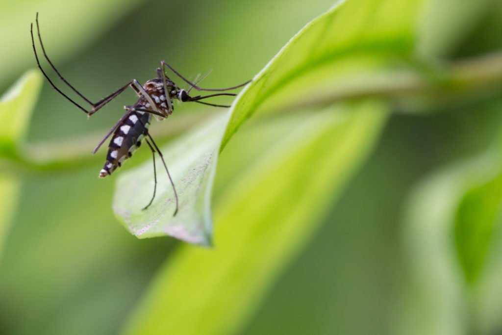
Aedes aegypti Mosquito. Close up a Mosquito Mosquito on leaf,Mosquito Vector-borne diseases,Chikungunya.Dengue fever.Rift Valley fever.Yellow fever.Zika virus.
Aedes aegypti mosquito, the vector of Chikungunya


Signs and Symptoms

Signs and Symptoms of Chikungunya develop after a 2-12 day incubation period. Cases vary in severity, and asymptomatic infection may occur. The rate of asymptomatic cases is estimated to be between 4% and 28%.

Cases often begin with an abrupt onset of fever. Polyarthralgia occurs in 70% of cases, usually involving small joints. Swelling of joints may also occur, typically without fluid accumulation. In greater than 50% of cases, a maculopapular rash on the palms, soles, limbs, torso, and/or face is present. This rash may progress to desquamation. Fever generally resolves within one week, but joint pain may persist for months. Sometimes, a “saddle-back fever curve” is seen, with fever resolving and then returning. Moderate to severe lymphopenia is often noted. Thrombocytopenia, leukopenia, elevated liver enzymes, anemia, and elevated creatinine may also be observed.

Facial and neck erythema and conjunctival suffusion may be noted. Headache, photophobia, retro-orbital pain, pharyngitis, nausea, and vomiting can occur. Sometimes, pneumonia and dry cough are seen. Pruritus is common. Patients may complain of exhaustion and insomnia. Symptoms of Chikungunya can persist from one week to several months. Residual chronic joint pain may continue in some cases. Chronic disease is more common in older patients and patients with prior rheumatological disease.

Chikungunya can also cause neurological and ophthalmologic complications. Eye involvement may include retinitis, retinal detachment, optic neuritis, uveitis, dendritic lesions, and Fuchs heterocyclic iridocyclitis. Neurological manifestations can include altered mental function, encephalitis, seizures, myelopathy, Guillain-Barré syndrome, bulbar palsy, acute flaccid paralysis, focal neurological deficit, and sudden sensorineural hearing loss.

Additional rare complications of Chikungunya include hemorrhagic syndrome, cardiovascular shock, arrhythmias, myopericarditis, renal failure, rhabdomyolysis, and thrombocytopenic purpura.

Children with Chikungunya are more likely to experience neurological and dermatological symptoms, and less likely to have arthralgia. Transplacental transmission of the virus can occur and may result in neonatal encephalopathy, neonatal respiratory distress, sepsis, necrotizing enterocolitis, and cardiologic complications. Infants who become infected during the perinatal period may experience fever, rash, peripheral edema, lymphopenia, and thrombocytopenia. Congenital and perinatal infections are associated with poor neurodevelopmental outcomes. Transmission of Chikungunya via breastfeeding has not been noted.

Fatalities from Chikungunya are rare, occurring in about 1 per 1,000 cases. Fatalities are more common in newborns and individuals with multiple medical comorbidities. The use of NSAIDs prior to hospitalization is associated with an increase in disease severity. Infection with Chikungunya is likely to protect against future disease. 


Diagnosis and Treatment

A diagnosis of Chikungunya should be considered in individuals living in – or having traveled to – areas with known outbreaks presenting with acute onset of fever and joint pain. Dengue fever and Zika virus infection should be considered in a differential diagnosis of Chikungunya, as they are also carried by Aedes species mosquitoes and may present with similar signs and symptoms.

PCR, serology, and viral culture can be used for laboratory confirmation of Chikungunya. Chikungunya is classified as a biosafety level-3 pathogen, and samples should be handled accordingly. Blood-borne transmission from patients to healthcare workers and laboratory personnel has been documented.

Patients with Chikungunya are treated with supportive care, including hydration and pain management. It is important to prevent mosquito bites during the first week of illness, in order to prevent additional transmission.


Chikungunya outbreaks

Between 1952 and 2013, Chikungunya virus outbreaks were identified in Africa, Asia, Europe, and the Indian and Pacific Oceans. In 2013, cases were first identified in the Americas and nations of the Caribbean, and today the majority of cases occur in these locations – where populations have no preexisting immunity.

Over the past decade, the countries that reported most cases of Chikungunya have included Haiti, Dominican Republic, Guadeloupe, Martinique, El Salvador, Honduras, Nicaragua, Columbia, Bolivia, Brazil, Ethiopia, Chad, India, Laos, and French Polynesia. If you have a GIDEON account, click to explore Chikungunya Outbreak Map.

Between 2004 and 2006, an outbreak of Chikungunya that began in Kenya resulted in 500,000 cases in countries of the Indian Ocean, including one-third of the population of La Reunion Island. This outbreak spread to India, where almost 1.5 million people were infected. Ongoing outbreaks have been occurring in Brazil since 2014, with over 300,000 cases occurring in 2016. It is thought that a mutation occurred around 2005 that enabled the virus to survive in Aedes albopictus; and that having this additional species as a vector has fueled recent outbreaks.

Local transmission was reported for the first time in Europe in 2007, with 197 cases occurring in north-eastern Italy. The source of this outbreak was traced to a single individual who had returned from India with the infection. A second outbreak occurred in Europe in 2014, centered mainly in France and the UK and resulting in about 1500 cases.

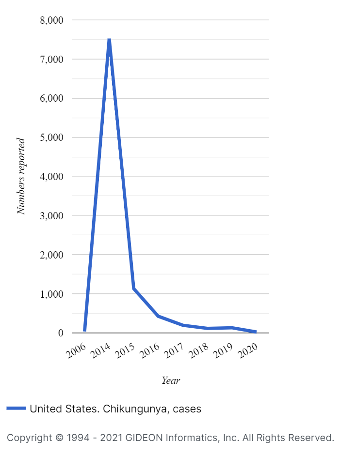
Chikungunya cases in United States, 2006 - 2020, GIDEON graph


In 2014, local transmission of the Chikungunya virus was identified in the territories of the United States for the first time, with 4,659 cases occurring between American Samoa, Puerto Rico, the U.S. Virgin Islands, and Florida. Since that time, the rate of local transmission in the United States has decreased each year, with 179 cases occurring in 2016, 8 cases in 2018, and no cases in 2020.


Prevention

There is currently no vaccine to prevent Chikungunya. The CDC recommends the use of the Environmental Protection Agency (EPA)-registered insect repellents when traveling to areas with outbreaks. Wearing long sleeves and pants can also reduce transmission, as can sleeping in places with air conditioning and window and door screens. The CDC also recommends using 0.5% permethrin to treat clothing and gear to repel mosquitos.

During outbreaks, measures should be taken to control mosquito populations by reducing both natural and artificial water-filled habitats where they may breed. Any items that may hold water, such as pools, buckets, planters, and trash containers, should be regularly emptied and cleaned.


Did you like this article? Share it on social media!

Check out more of our latest content here


This blog was written using data from the GIDEON database, CDC, and WHO.

Hospital-Acquired Infections

by Dr. Jaclynn Moskow

Hospital-acquired infections, also known as “healthcare-associated infections” or “nosocomial infections,” refer to infections that were not present before seeking medical care and were acquired in a healthcare setting. Hospital-acquired infections can be contracted in hospitals, ambulatory clinics, surgical centers, nursing homes, long-term care facilities, dialysis centers, and diagnostic laboratories. 

Hospital setting: male nurse pushing stretcher gurney bed in hospital corridor with doctors & senior female patient

Hospital-acquired infections are defined by symptoms presenting 48-or-more hours after hospital admission, within three days of discharge, or 30 days postoperatively (1). The vast majority of hospital-acquired infections are caused by bacteria, and the propagation of these infections is worsened by the increasing presence of multi-drug resistant bacterial strains.


Prevalence of hospital-acquired infections

In the United States, approximately 1 in 25 hospitalized patients will contract an infection (2). Data collected by the Centers for Disease Control and Prevention identified an estimated 1.7 million hospital-acquired infections in the United States during 2002, resulting in 99,000 associated deaths (3).

Estimates from the UK place the prevalence of hospital-acquired infections at approximately 1-in-10 patients (1). In developing nations, the prevalence is higher and may occasionally exceed 25% (4).

CDC data show that urinary tract infections make up approximately 36% of all hospital-acquired infections in the ICU, surgical site infections 20%, pneumonias 11%, bloodstream infections 11%, and other infections 22% (3).


Risk Factors

Immunocompromised individuals, such as those undergoing chemotherapy, are at an increased risk for hospital-acquired infection. Geriatric patients are also at increased risk, as are those with multiple medical comorbidities. The incidence of hospital-acquired infections increases as the length of hospital stay increases. Patients in the ICU, receiving mechanical ventilator support, undergoing surgery, and having indwelling devices are also at increased risk.

One large study that examined 231,459 patients across 947 hospitals in Europe found that 19.5% of patients in the ICU experienced at least one hospital-acquired infection (5).


Catheter-Associated Urinary Tract Infections (CAUTI)

Catheter-associated urinary tract infections are the most common forms of hospital-acquired infection. Approximately 75% of all UTIs contracted in the hospital are associated with catheter use, and the most important risk factor for developing a catheter-associated urinary tract infection is prolonged catheter use (6). Common pathogens identified in catheter-associated urinary tract infections include Escherichia coli, Enterococcus species, Staphylococcus aureus, Pseudomonas aeruginosa, Proteus mirabilis, Klebsiella pneumoniae, Morganella morganii, and Candida albicans. Some organisms, including Pseudomonas and Proteus, can form biofilms around catheters.


Surgical Site Infections (SSI)

Surgical site infections occur postoperatively in the skin, internal organs, or implanted materials involved in the surgery. Diabetic patients are at an increased risk of developing surgical site infections. The incidence of surgical site infections increases as procedure duration increases and the use of antimicrobial prophylaxis decreases the risk of such infections. Common causes of surgical site infections include Staphylococcus aureus (including MRSA), coagulase-negative Staphylococcus, Escherichia coli, Enterococcus faecalis, Pseudomonas aeruginosa, Klebsiella pneumoniae, and Acinetobacter baumannii. In developed nations, between 2-5% of all patients who undergo surgery develop a surgical site infection; and in developing nations, between 12%-39% do (4).


Hospital-Acquired Pneumonia (HAP) and Ventilator-Associated Pneumonia (VAP)

The Infectious Diseases Society of America (IDSA) defines hospital-acquired pneumonia as “pneumonia that occurs 48 hours or more after admission to the hospital and did not appear to be incubating at the time of admission”; and defines ventilator-associated pneumonia as “pneumonia that develops more than 48 to 72 hours after endotracheal intubation.” Common bacterial causes of both hospital-acquired pneumonia and ventilator-associated pneumonia include Staphylococcus aureus (including MRSA), Streptococcus pneumoniae, Haemophilus influenzae, Escherichia coli, Pseudomonas aeruginosa, and Klebsiella pneumoniae. Common viral causes include rhinovirus, parainfluenza virus, influenza virus, respiratory syncytial virus, and coronavirus. 

The incidence of ventilator-associated pneumonia in patients who require mechanical ventilation for more than 48 hours is estimated at 25-to-30% (7).


The male patient with intravenous catheter. Central Line-Associated Bloodstream Infection (CLABSI) is one of the types of hospital-acquired infections

Central Line-Associated Bloodstream Infection (CLABSI)

Central line-associated bloodstream infections occur at the site of central venous catheters. The mortality rate for central line-associated bloodstream infections is between 12% and 25% (8). Common causes of central line-associated bloodstream infections include coagulase-negative Staphylococci, Staphylococcus aureus (including MRSA), Enterobacte, Klebsiella pneumoniae, and Candida albicans. Central lines can be placed in the neck, chest, arm, or groin. The use of femoral-site lines is associated with an increased risk of infection and is no longer recommended (9). Antibiotic lock therapy can reduce the incidence of central line-associated bloodstream infections.


Clostridium Difficile Infections (CDI)

An estimated 12.1% of all hospital-acquired infections are caused by Clostridium difficile, making Clostridium difficile the most common cause of hospital-acquired infections (10). Approximately 75% of all Clostridium difficile infections are hospital-acquired (11), and an estimated 2.3% of all US hospital costs are related to these infections (12). Click to see how you can use Gideon to explore Clostridium difficile. 


Hospital-Acquired COVID-19

The incidence of hospital-acquired COVID-19 remains unknown. A meta-analysis of studies examining COVID-19 cases in China found that 44% of cases were likely to have originated from a healthcare setting (13). A hospital in South Africa reported that a single case led to six major outbreak clusters in several hospital wards, a nursing home, and a dialysis unit. Ultimately this episode resulted in 135 infections and 15 deaths (14). Up to 1-in-4 cases of COVID-19 in the UK are likely to have been hospital-acquired (15).

In contrast, a recent study from the United States suggests that hospital-acquired COVID-19 is actually quite uncommon when rigorous infection-control measures are followed. This study looked at all patients admitted to Brigham and Women’s Hospital in Boston, Massachusetts, between March 7 and May 30, 2020. They determined that of 697 COVID-19 diagnoses, only two were hospital-acquired, including one case that likely resulted from a visit by a pre-symptomatic spouse (16).

The World Health Organization estimates that healthcare workers may comprise as many as one-in-seven COVID-19 cases (17), reflecting a high incidence of hospital-acquired disease. The CDC is not currently collecting data on hospital-acquired COVID-19, as hospitals are required to report to the U.S. Department of Health and Human Services. 


Did you like this article? Share it on social media!

Check out more of our latest content here


References 

(1) Inweregbu, K., Dave, J. and Pittard, A., 2005. Nosocomial infections. Continuing Education in Anaesthesia Critical Care & Pain, 5(1), pp.14-17.

(2) Magill SS, Edwards JR, Bamberg W, et al., 2014. Emerging Infections Program Healthcare-Associated Infections and Antimicrobial Use Prevalence Survey Team. Multistate point-prevalence survey of healthcare-associated infections. N Engl J Med, 27;370(13), pp. 1198-208.

(3) Klevens, R., Edwards, J., Richards, C., et al., 2007. Estimating Health Care-Associated Infections and Deaths in U.S. Hospitals, 2002. Public Health Reports, 122(2), pp.160-166.

(4) Allegranzi, B. and Pittet, D., 2007. Healthcare-Associated Infection in Developing Countries: Simple Solutions to Meet Complex Challenges. Infection Control & Hospital Epidemiology, 28(12), pp.1323-1327. 

(5) European Centre for Disease Prevention and Control, 2013. Point-prevalence survey of healthcare-associated infections and antimicrobial use in European acute care hospitals. Stockholm: EDC.

(6) Cdc.gov. 2021. Catheter-associated Urinary Tract Infections (CAUTI) | HAI | CDC. [online] Available at: https://www.cdc.gov/hai/ca_uti/uti.html.

(7) Cornejo-Juárez, P., González-Oros, I., Mota-Castañeda, P., Vilar-Compte, D. and Volkow-Fernández, P., 2020. Ventilator-associated pneumonia in patients with cancer: Impact of multidrug resistant bacteria. World Journal of Critical Care Medicine, 9(3), pp.43-53.

(8) Dumont, C. and Nesselrodt, D., 2012. Preventing central line-associated bloodstream infections CLABSI. Nursing, 42(6), pp.41-46. 

(9) Palmer, E., 2021. Avoiding the femoral vein in central venous cannulation: an outdated practice. [online] Acphospitalist.org. Available at: https://acphospitalist.org/archives/2018/08/perspectives-avoiding-the-femoral-vein-in-central-venous-cannulation-an-outdated-practice.htm.

(10) Monegro, A., Muppidi, V. and Regunath, H., 2020. Hospital Acquired Infections. StatPearls, [online] Available at: https://www.ncbi.nlm.nih.gov/books/NBK441857/.

(11) Louh, I., Greendyke, W., Hermann, E., e al., 2017. Clostridium Difficile Infection in Acute Care Hospitals: Systematic Review and Best Practices for Prevention. Infection Control & Hospital Epidemiology, 38(4), pp.476-482.

(12) Jump, R., 2013. Clostridium difficile infection in older adults. Aging health, 9(4), pp.403-414.

(13) Zhou, Q., Gao, Y., Wang, X., et al., 2020. Nosocomial infections among patients with COVID-19, SARS and MERS: a rapid review and meta-analysis. Annals of Translational Medicine, 8(10), pp.629-629.

(14) Lessells, R., Moosa, Y. and de Oliviera, T., 2020. Report into a nosocomial outbreak of coronavirus disease 2019 (COVID‐19) at Netcare St. Augustine’s Hospital. [online] Available at: https://www.krisp.org.za/manuscripts/.

(15) Discombe, M., 2021. Covid infections caught in hospital rise by a third in one week. [online] Health Service Journal. Available at: https://www.hsj.co.uk/patient-safety/covid-infections-caught-in-hospital-rise-by-a-third-in-one-week/7029211.article. 

(16) Rhee, C., Baker, M., Vaidya, V., et al., 2020. Incidence of Nosocomial COVID-19 in Patients Hospitalized at a Large US Academic Medical Center. JAMA Network Open, 3(9), p.e2020498.

(17) Nebehay, S., 2021. One in 7 reported COVID-19 infections is among health workers, WHO says. [online] U.S. Available at: https://www.reuters.com/article/us-health-coronavirus-who-healthworkers/one-in-7-reported-covid-19-infections-is-among-health-workers-who-says-idUSKBN2681TR?il=0. 

‘It’s not just COVID-19’ – Dr. Berger on Outbreak News Today podcast

HazMat team in protective suits decontaminating public transport, bus interior during virus outbreak

The global pandemic caused by COVID-19 has rightly taken center stage in media and scientific journals but overshadowed other concerning outbreaks that could do with some attention. GIDEON co-founder Dr. Stephen A. Berger has been speaking with Outbreak News Today to discuss the diseases that are flying under the radar in the media but are still being tracked and reported by GIDEON. 

Listen to the podcast or watch a video recording here.

In 2020, significant outbreaks of Cholera in Yemen, Dengue in Brazil, and neighboring South American countries have been recorded in addition to the COVID-19 pandemic. Numerous diseases such as Ebola, Lassa fever, Chikungunya, Plague, and Monkeypox have broken out in regions of Africa and Asia in recent years as well. Ebola and Monkeypox have proved a persistent threat in the Democratic Republic of Congo, with thousands of cases in the last couple of years alone. Meanwhile, Lassa fever cases in Nigeria in 2020 were the highest recorded by any country in history (nearly 7,000). The disease spreads through rodents, leaving many of its surviving victims deaf.

These and other diseases have historically been considered tropical or exotic and don’t trouble the western population too much, however, the spread of COVID-19 has proven that diseases can and will spread given the opportunity. For instance, Monkeypox, Plague, and West Nile Fever have all had outbreaks within the US in the past. 

Tune in to Outbreak News Today and hear from Dr. Berger and Robert Herriman on this timely subject.

Haven’t got a GIDEON account yet? Sign up for a free trial here.


Did you like this article? Share it on social media!

Check out more of our latest content here

The Gut Microbiome and Its Role in Health and Disease

by Dr. Jaclynn Moskow

Intestinal bacteria, Gut microbiome helps control intestinal digestion and the immune system, Probiotics are beneficial bacteria used to help the growth of healthy gut flora
The gut microbiome helps control intestinal digestion and the immune system.


It is difficult to overstate the importance and complexity of the gut microbiome. Humans live in symbiosis with hundreds (and possibly thousands) of species of bacteria (1). Additionally, archaea, fungi, viruses, and protozoa are also present in our gut. In fact, only about 10% of the cells within our bodies are “ours” and contain human DNA. The remaining 90% of cells we carry with us are microbial. The exact makeup of the gut microbiome varies greatly from individual to individual and is influenced by variables that include diet, exercise, medication use, sleep, stress, hormonal changes, aging, and disease. Associations have been found between the composition of the microbiome and obesity, diabetes, hypertension, heart disease, autoimmune disorders, allergies, mood disorders, and more.


Why Do We Have a Gut Microbiome?

The bacteria in our gut participate in the digestion, absorption, and metabolism of proteins and carbohydrates and in the breakdown of endogenous intestinal mucus. They also synthesize vitamin K2 and various B group vitamins; and they influence the development of gut-associated lymphoid tissues and the development of cells of the immune system (2), and serve to limit the colonization of pathogenic bacteria. The majority of these bacteria are anaerobic. Common genera include Escherichia, Bifidobacterium, Lactobacillus, and Enterococcus.


The Gut Microbiome and COVID-19

A recent study examined the connection between the gut microbiome and COVID-19. Researchers found that patients hospitalized for COVID-19 had an increase in certain bacterial species and a decrease in others when compared to a control group, even after antibiotic use was accounted for (19). They found a negative correlation between disease severity and concentrations of Faecalibacterium prausnitzii and Eubacterium rectale. Patients were monitored for 30 days post-recovery, and the observed changes persisted. The researchers postulated that these changes may contribute to the persistence of symptoms and multi-system inflammation that is sometimes seen with patients who have recovered from COVID-19.


Gut flora vector illustration. Flat tiny gastrointestinal microbe person concept. Abstract digestive stomach living organisms for healthy life. Lactobacilli, coli and intestinal system environment.


The Gut Microbiome and Obesity

In recent years, many studies have examined associations between the gut microbiome and obesity. When germ-free mice are colonized with gut bacteria from obese mice, they gain weight; but when they are colonized with gut bacteria from lean mice, they do not gain weight (3). Mice also gain weight when they are colonized with bacteria from obese humans. In a discordant twin study, colonization from obese twins caused mice to gain weight while colonization from their lean siblings did not (4). Some believe the ratio of Bacteroidetes to Firmicutes may play a significant role in obesity. One study found that as obese individuals lose weight, the concentration of Bacteroidetes increases (5). Furthermore, genetically obese mice contain a higher proportion of Firmicutes than thin mice consuming the same diet, and thin mice contain more Bacteroidetes than obese mice consuming the same diet (6). When researchers employed machine learning to study this topic, they concluded that the association between the Bacteroidetes / Firmicutes ratio and obesity is relatively weak and that existing studies lack significant sample sizes (7). The science is far from settled.


The Gut Microbiome and Diabetes 

Studies have also investigated the link between the gut microbiome and diabetes. Some speculate that in individuals who are genetically susceptible to type 1 diabetes, it is ultimately a shift in the gut microbiome that triggers the onset (8).  The gut microbiome of children with type 1 diabetes has been found to be less diverse than that of children without the disease (9). A recent review of 42 studies that examined the gut microbiome and type 2 diabetes found Bifidobacterium, Bacteroides, Faecalibacterium, Akkermansia, and Roseburia to be negatively associated with type 2 diabetes; and Ruminococcus, Fusobacterium, and Blautia to be positively associated (10). Other work has shown that when individuals with metabolic syndrome were given fecal transplants from healthy donors, insulin-resistance improved (11).


The Gut Microbiome, Hypertension, and Cardiovascular Disease

The ratio of Bacteroidetes to Firmicutes has also been implicated in hypertension. Consuming milk fermented with Lactobacilli can lower blood pressure in some cases, and Lactobacilli produce peptides that can inhibit ACE1 (12).  The same bacterial species found within the atherosclerotic lesions of individuals with cardiovascular disease are found in their gut (13). Additionally, Akkermansia muciniphila may have a cardioprotective effect. Researchers observed that when mice were fed a Western diet, they experienced a decrease in Akkermansia muciniphila and an increase in atherosclerotic lesions. When these same mice were recolonized with Akkermansia muciniphila, a reversal in atherosclerotic lesions was observed (14). 


The Gut Microbiome, Autoimmune Disorders, and Allergies

Components of the gut microbiome may be involved in eliciting or quelling immune responses that lead to the development of autoimmune disorders and allergies. Antibodies directed against a yeast species, Saccharomyces cerevisiae,  have been found in patients with rheumatoid arthritis, systemic lupus erythematosus, antiphospholipid syndrome, and Crohn’s Disease (15). Individuals with these conditions show an increase in the numbers of certain bacterial species and a decrease in other species –  as do individuals with multiple sclerosis, Sjögren’s syndrome, and celiac disease. The ratio of Clostridium difficile to Bifidobacterium in infants has been associated with food and aero-allergies, and high levels of fecal Escherichia coli in infants are associated with IgE-mediated eczema (16).


The Gut Microbiome and Neuropsychiatric Disorders

The central nervous system and enteric nervous system (together known as the gut-brain axis) are both influenced by the gut microbiome. Bacteria in the gut can directly secrete neurotransmitters, including serotonin, dopamine, norepinephrine, GABA, and histamine. Several studies have shown that patients with bipolar and major depressive disorder have an increase in Actinobacteria and Enterobacteriaceae, and a decrease in Faecalibacterium (17). Mice treated with Lactobacillus rhamnosus have reduced anxiety/depression-like behavior and altered expression of GABA receptors (18). Differences in microbiome composition have also been noted in patients with schizophrenia, Parkinson’s disease, and an autism spectrum disorder.


Fecal microbiota transplant (FMT) stool transferring bacteria microbes
Fecal microbiota transplantation (FMT)


Fecal Microbiota Transplantation and Clostridium Difficile Colitis

Fecal microbiota transplantation (FMT) is currently being used as a treatment for Clostridium difficile colitis. In fact, FMT is more effective than vancomycin at treating recurrent Clostridium difficile colitis. Most commonly, FMT is performed via colonoscopy. Nasoduodenal tubes, nasogastric tubes, and enemas can also be used. FMT made headlines in 2019 when a transplant recipient died, and several others became seriously ill, after becoming colonized with multi-drug resistant Escherichia coli. This led the FDA to recommend new safety measures for FMTs, including screening donors for risk factors associated with carrying multi-drug-resistant organisms and testing all donor stools for such organisms.


Optimizing the Microbiome

In many regards, studying the gut microbiome often leads to more questions than answers. When a change in bacterial levels is observed in a disease state, it is sometimes difficult to know whether that change contributed to the disease state or merely resulted from it. Anyone who seeks to convince you that they know the perfect solution to optimizing gut health is misleading you. While a host of food products and health supplements are touted to enhance the gut microbiome, in most cases the details of this “enhancement” are not defined. As additional studies are conducted, we will gain a better understanding of this vast topic and will likely see an increase in the utilization of fecal transplants in treating various diseases.


Did you like this article? Share it on social media!

Check out more of our latest content here


References

(1) Almeida A, Mitchell AL, Boland M, et al. A new genomic blueprint of the human gut microbiota. Nature. 2019 Apr;568(7753):499-504. Available: 10.1038/s41586-019-0965-1.

(2) Wu HJ, Wu E. The role of gut microbiota in immune homeostasis and autoimmunity. Gut Microbes. 2012 Jan-Feb;3(1):4-14. Available: 10.4161/gmic.19320.

(3) Turnbaugh PJ, Ley RE, Mahowald MA, et al. An obesity-associated gut microbiome with increased capacity for energy harvest. Nature. 2006 Dec 21;444(7122):1027-31. Available: 10.1038/nature05414. 

(4) Ridaura VK, Faith JJ, Rey FE, et al. Gut microbiota from twins discordant for obesity modulate metabolism in mice. Science. 2013 Sep 6;341(6150):1241214. Available: 10.1126/science.1241214.

(5) Ley RE, Turnbaugh PJ, Klein S, Gordon JI. Microbial ecology: human gut microbes associated with obesity. Nature. 2006 Dec 21;444(7122):1022-3. Available: 10.1038/4441022a.

(6) Ley RE, Bäckhed F, Turnbaugh P, et al. Obesity alters gut microbial ecology. Proc Natl Acad Sci U S A. 2005 Aug 2;102(31):11070-5. Available: 10.1073/pnas.0504978102.

(7) Sze MA, Schloss PD. Looking for a Signal in the Noise: Revisiting Obesity and the Microbiome. mBio. 2016 Aug 23;7(4):e01018-16. Available: 10.1128/mBio.01018-16.

(8) Zheng P, Li Z, Zhou Z. Gut microbiome in type 1 diabetes: A comprehensive review. Diabetes Metab Res Rev. 2018 Oct;34(7):e3043. Available: 10.1002/dmrr.3043.

(9) Giongo A, Gano KA, Crabb DB, et al. Toward defining the autoimmune microbiome for type 1 diabetes. ISME J. 2011 Jan;5(1):82-91. Available: 10.1038/ismej.2010.92.

(10) Gurung M, Li Z, You H, et al. Role of gut microbiota in type 2 diabetes pathophysiology. EBioMedicine. 2020 Jan;51:102590. Available: 10.1016/j.ebiom.2019.11.051.

(11) Vrieze A, Van Nood E, Holleman F, et al. Transfer of intestinal microbiota from lean donors increases insulin sensitivity in individuals with metabolic syndrome. Gastroenterology. 2012 Oct;143(4):913-6.e7. Available: 10.1053/j.gastro.2012.06.031.

(12) Jose PA, Raj D. Gut microbiota in hypertension. Curr Opin Nephrol Hypertens. 2015 Sep;24(5):403-9. Available: 10.1097/MNH.0000000000000149.

(13) Tang WH, Kitai T, Hazen SL. Gut Microbiota in Cardiovascular Health and Disease. Circ Res. 2017 Mar 31;120(7):1183-1196. Available: 10.1161/CIRCRESAHA.117.309715.

(14) Li J, Lin S, Vanhoutte PM, et al. Akkermansia Muciniphila Protects Against Atherosclerosis by Preventing Metabolic Endotoxemia-Induced Inflammation in Apoe-/- Mice. Circulation. 2016 Jun 14;133(24):2434-46. Available: 10.1161/CIRCULATIONAHA.115.019645.

(15) De Luca F, Shoenfeld Y. The microbiome in autoimmune diseases. Clin Exp Immunol. 2019 Jan;195(1):74-85. Available: 10.1111/cei.13158.

(16) Pascal M, Perez-Gordo M, Caballero T, et al. Microbiome and Allergic Diseases. Front Immunol. 2018 Jul 17;9:1584. Available: 10.3389/fimmu.2018.01584.

(17) Huang TT, Lai JB, Du YL, et al. Current Understanding of Gut Microbiota in Mood Disorders: An Update of Human Studies. Front Genet. 2019 Feb 19;10:98. Available: 10.3389/fgene.2019.00098.

(18) Bravo JA, Forsythe P, Chew MV, et al. Ingestion of Lactobacillus strain regulates emotional behavior and central GABA receptor expression in a mouse via the vagus nerve. Proc Natl Acad Sci U S A. 2011 Sep 20;108(38):16050-5. Available: 10.1073/pnas.1102999108.

(19) Yeoh YK, Zuo T, Lui GC, et al. Gut microbiota composition reflects disease severity and dysfunctional immune responses in patients with COVID-19. Gut. 2021 Jan 11:gutjnl-2020-323020. Available: 10.1136/gutjnl-2020-323020.

GIDEON Sign In